Any given day in a ruminant slaughterhouse (2/2)
We continue with this second post dedicated to the lesions that were observed in condemned viscera from bovine carcasses.
In previous posts we discussed some of the most common lesions that can be found in swine and small ruminants.
Case 1
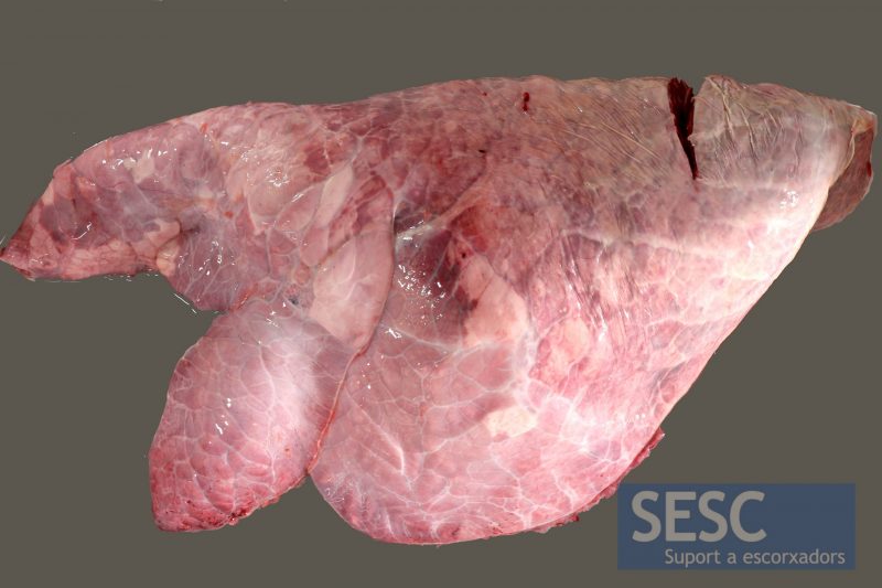
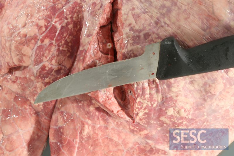
Bovine lung with a consolidation of the parenchyma of anteroventral distribution , this lesion corresponds to a suppurative bronchopneumonia.
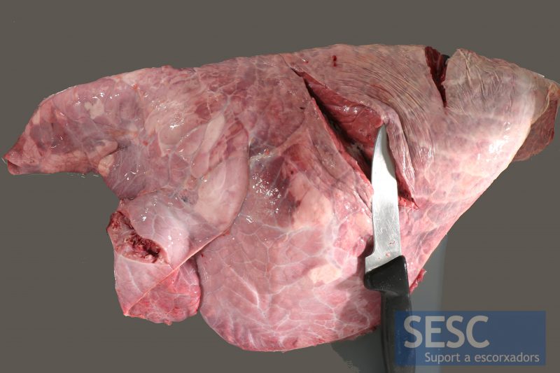
It is harder to see that in the lungs of small ruminants because the pleura in cattle is thicker, but the consistency of the anteroventral pulmonary parenchyma is increased and when sectioned we can see the presence of suppurative exudate (while the appearance of the parenchyma - top cut with the knife - is normal).
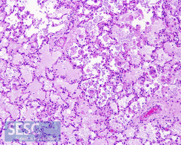
Presence of alveolar edema and inflammatory cells (macrophages and neutrophil polymorphonuclear leukocytes).
Case 2
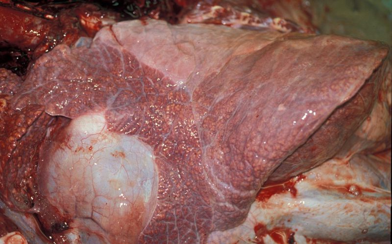
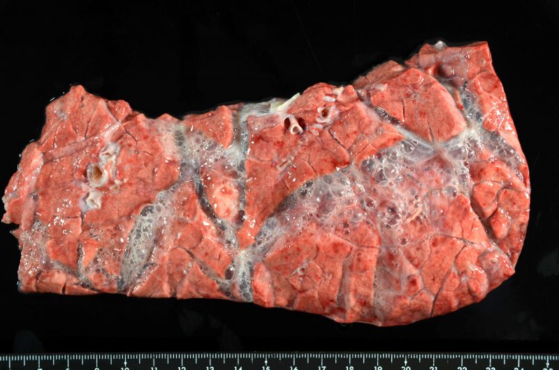
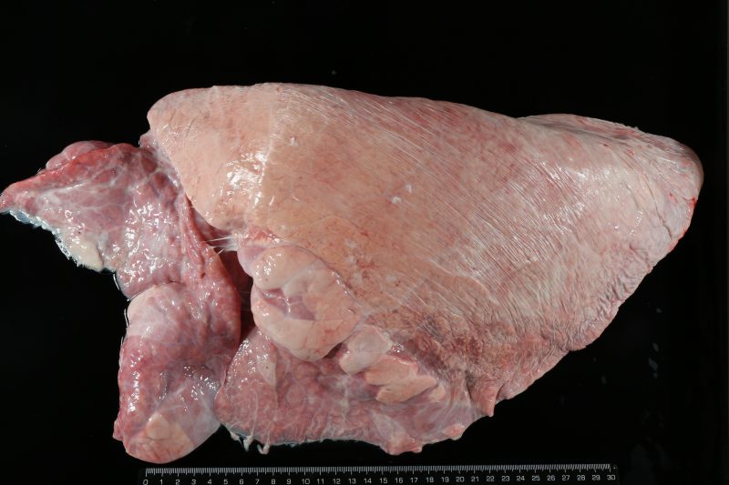
A more chronic example of suppurative bronchopneumonia. The rough appearace of the pulmonary parenchyma is due to the dilatation of the bronchi due to the presence of suppurative exudate (bronchiectasis). This image is not from a slaughterhouse but from the necropsy room, SDPV, UAB Veterinary Faculty.
Case 3
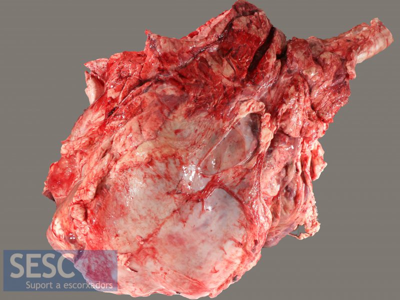
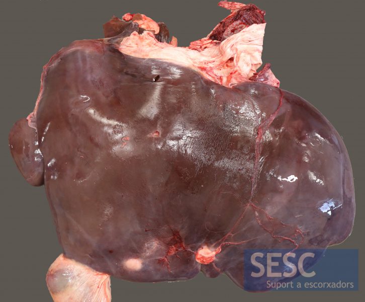
Bovine heart increased in size and with the pericardium adhered to epicardium.
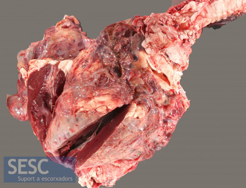
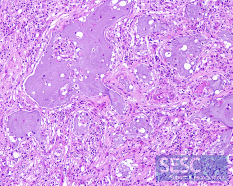
When sectioned we can observe that it is a fibrous pericarditis. Swelling and presence of fibrous adhesions between the epicardium and pericardium. This is a chronification of a fibrinous exudate. The cause is unknown, one possibility is a traumatic reticulopericarditis.
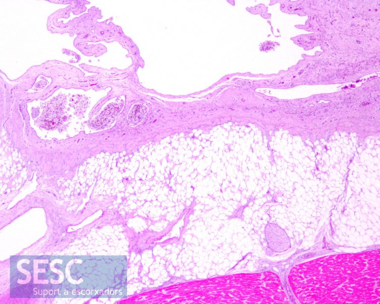
Markedly swollen epicardium, with infiltration of fibrous connective tissue and adipose tissue and villous projections (above).
Case 4
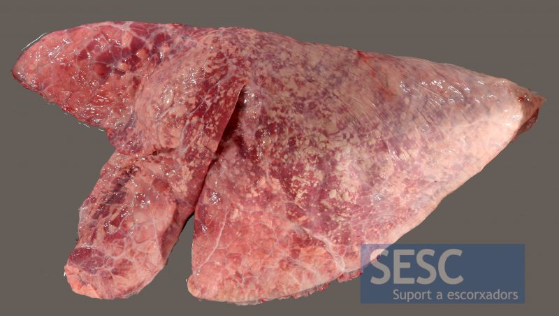
Bovine lung with darkened areas and witish areas of dorsoposterior distribution.
When sectioned, it can be observed that the coloration change affects only the superficial layer of the pulmonary parenchyma, the rest has a normal appearance. It is another case of atelectasis (collapse of the alveoli). The lack of air inside the alveoli gives it a darker color. Also, the whitest areas correspond to air -filled alveoli. It is not a lesion per se.
Case 5
Bovine lung section with presence of air (bubbles) in the interlobular septa. It is an interstitial emphysema caused most probably by spasmodic dilatations of the thoracic cavity during the sacrifice that increase the air pressure within the alveoli, it is not an in vivo lesion, but a mechanical alteration that causes the air to exit from the alveoli to the interstitial space. Image: SDPV, UAB Veterinary Faculty.
Case 6
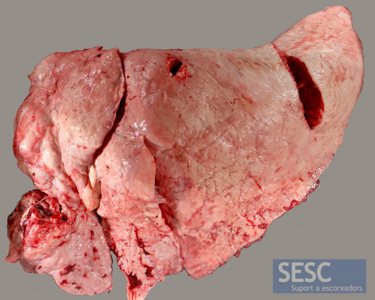
Bovine lung with an anteroventral area of consolidation of the pulmonary parenchyma (in the lower left corner of the image).
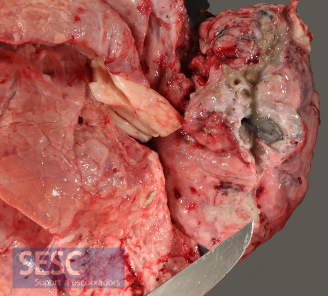
When sectioned the lesion gives off a strong putrid odor and the appearance is cavernous and of greenish-gray coloration. It is a gangrenous pneumonia. The appearance and the smell are caused by the proliferation of bacteria. One possible cause is the deviated swallowing of food material, known as aspiration pneumonia.
Histology showed fibrosis, inflammatory infiltrate rich in macrophages and neutrophil polymorphonuclear leukocytes, pulmonary parenchyma necrosis and secretion with abundant bacteria and cell detritus.
Case 7
Bovine liver with multiple abscesses.
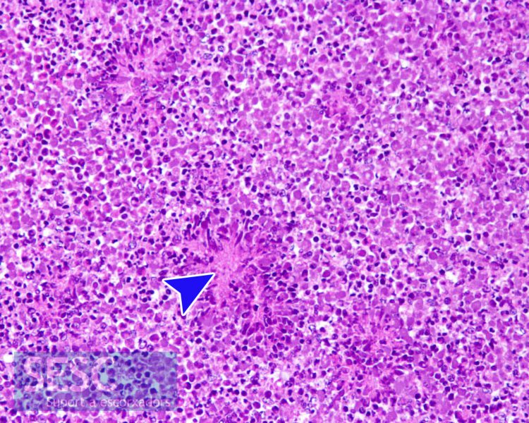
Abscesses are formed by accumulation of neutrophil polymorphonuclear leukocytes in different degeneration phases, sometimes surrounding bacterial colonies (arrowhead).
Case 8
Bovine lung with suppurative bronchopneumonia (notice the darkened areas of anteroventral consolidation). On the limit between the lesioned area with the normal pulmonary parenchyma some whitish areas of parenchyma can be observed. In the past, this finding was described as vicarious emphysema, this nomenclature refers to the vicar (substitute for the bishop) since it was believed that they were lung areas that compensated for the lack of functionality of the injured area. This is NOT correct, it simply consists of areas with a lack of alveolar collapse, probably because the presence of inflammatory exudation which has prevented the air from flowing out of the alveoli. Image: SDPV, UAB Veterinary Faculty.
