Any given day in a ruminant slaughterhouse (1/2)
Last Friday, July the 12th we organized the 2nd workshop for the identification and description of lesions for official meat inspectors.
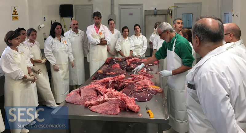
The course was given by Dr. Mariano Domingo UAB, IRTA-CReSA, of the Dr. Jorge Martínez UAB iRTA-CReSA and Dr. Alberto Marco UAB. Twenty-one people attended who scored the activity very positively.
Case 1
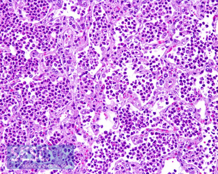
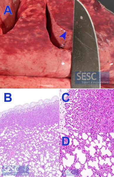
Small ruminant lung with areas of consolidation of the pulmonary parenchyma of cranio-ventral distribution, typical image of suppurative bronchopneumonia, of bacterial etiology.
A: On the left, pulmonary parenchyma is healthy and, on the right, suppurative exsudate-filled alveoli. B: Detail of the alveoli full of neutrophilic polymorphonuclear leukocytes (PMNN). C: Detail of a bronchiole filled with purulent inflammatory exudate.
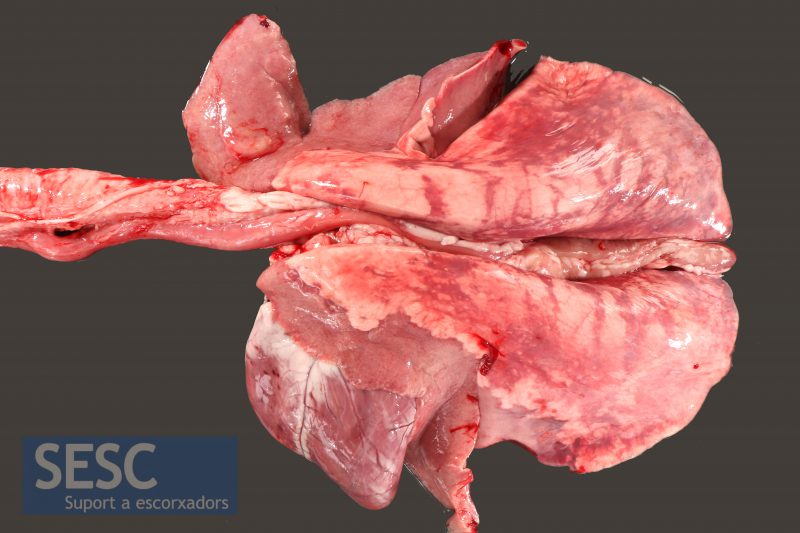
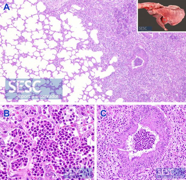
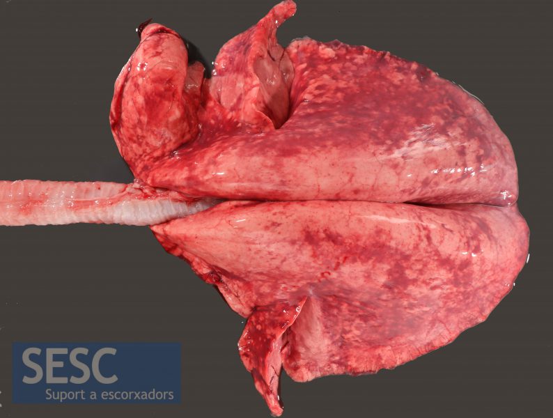
Case 2

Small ruminant lung with multiple white nodular lesions.
WHen sectiones the lesions appear necrotic and are encapsulated.
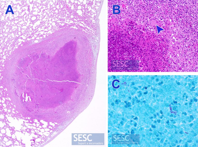
A: Histologically the lesions consisted of encapsulated nodules with a piogranulomatous inflammatory infiltrate compatible with tuberculosis. B: Detail of the inflammatory infiltrate with the presence of Langhans multinucleated giant cells (arrowhead). C: Ziehl Neelsen's staining showed the presence of mycobacteria on the lesion. This case is a good example of why all the granulomatous lesions must to be sent to the laboratory not to miss any new outbreaks of this zoonosis.
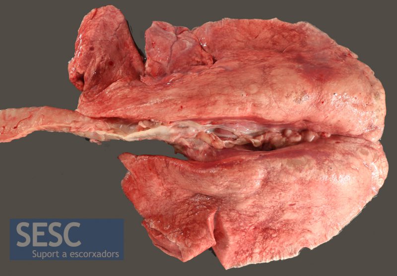
Case 3
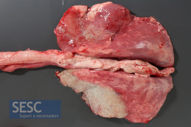
Small ruminant lung with anteroventral consolidation areas (suppurative bronchopneumonia) and presence of fibrous material in the pleura (fibrous pleuritis).
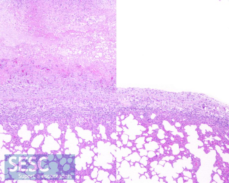
To the right, normal pleura, and to the left, thickened pleura with fibrinous secretion in the phase of organization with connective tissue and vascularization.
In this case, suppurative exudates were also observed inside the alveoli and bronchioles.
Case 4
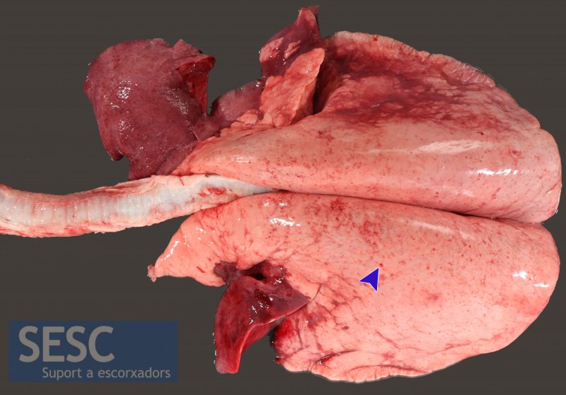
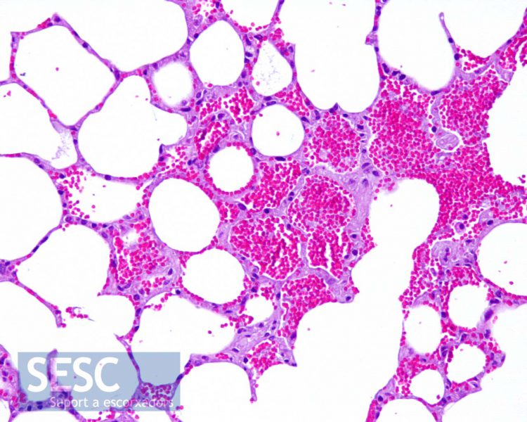
Small ruminant lung with extensive reddened areas.
A: When sectioning it is observed that the darkening only affects the more superficial layer of the parenchyma (arrowhead). B: Histologically, an atelectasis or collapse of the superficial alveoli is observed. No presence of inflammatory cells (C). D: The underlying pulmonary parenchyma is normal. It is thus an artifact and not a lesion.
Case 5
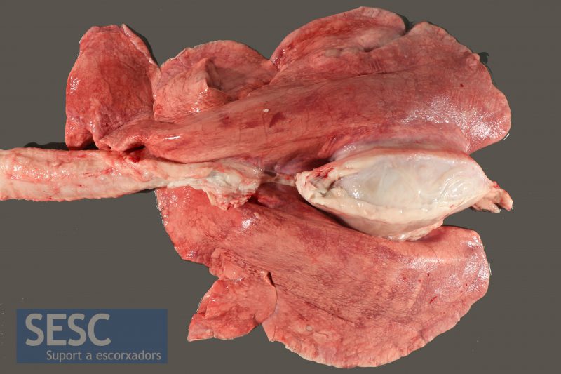
Another small ruminant lung with suppurative bronchopneumonia. In this case, the presence of fibrinous exudate (arrowhead: yellowish material, friable, easy to peel off and a texture similar to that of a french omelette) is found on the pleura: fibrinous pleuritis. It is a case similar to case 3 but more acute.
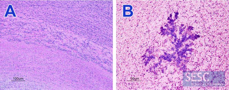
Case 6
Small ruminant lung with multiple lesions of gray coloration of dorso-caudal distribution.
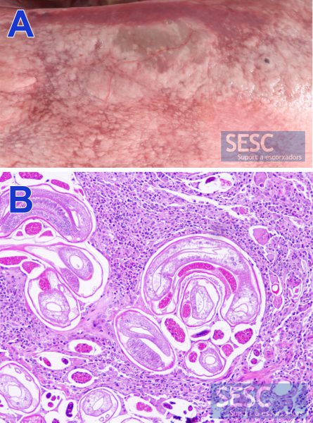
A: Detail of the lesions. B: Microscopically, they correspond to a granulomatous pneumonia with the presence of larvae of nematode parasites. This is a verminous pneumonia and usually presents this characteristic grayish appearance.
Case 7
This small ruminant lung also had suppurative bronchopneumonia and, in addition, multiple red spots with a haemorrhagic aspect (arrowhead) distributed on the dorsal aspect could be observed.
These spots should not be interpreted as petechiae (haemorrhages), but rather as aspiration of blood, which may occur during the cutting of the trachea. In histology, blood can be seen inside the alveoli. This alteration is very common in ritual sacrifices without stunning (hallal or kosher). This should not be confused with aspiration pneumonia that refers to when the animal has a deviant swallowing of food content trough the trachea which results in a very serious necrosis and inflammatory reaction.
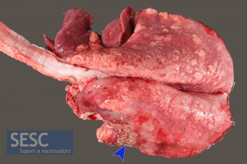
Case 8
Small ruminant lung with hyperplasia of the mediastinal lymph node and the presence of whitish nodules in the pulmonary parenchyma.
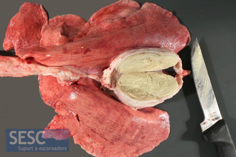
When sectioned it can be observed that it is a caseous lymphadenitis, with the characteristic concentric pattern of organization in onion layers. The causative agent is Corynebacterium pseudotuberculosis. These lesions are quite common and can be concomitant with a granulomatous pneumonia due to tuberculosis.
A: Histology shows overlapping layers of degenerated inflammatory cells (PMNN and macrophages). B: The Gram staining allows to demonstrate the colonies of Gram + bacteria that cause the lesion.
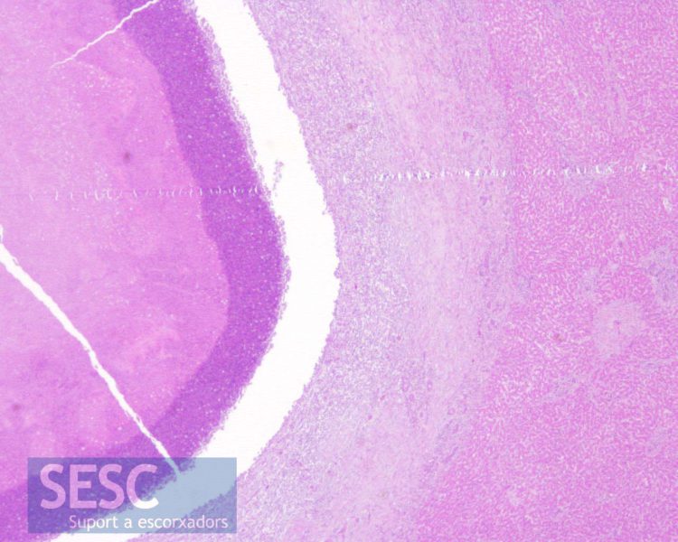
Case 9
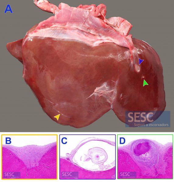
A: Small ruminant liver with multiple parasitic lesions corresponding to different stages of Cysticercus tenuicollis (Taenia hydatignea larval phase): viable cyst (blue arrowhead), superficial granuloma (green arrowhead) and migration path (yellow arrowhead). Histology shows the different lesions: B: fibrous scar in the case of the migration path. C: Vesicle with the scolex inside. D: Liver parasitic granuloma with mineralized necrotic center. This parasite should not be confused with a hidatidosis that presents intraparenchymal lesions and not these typical vesicles to serous lashes, such as C.tenuicollis.
Case 10
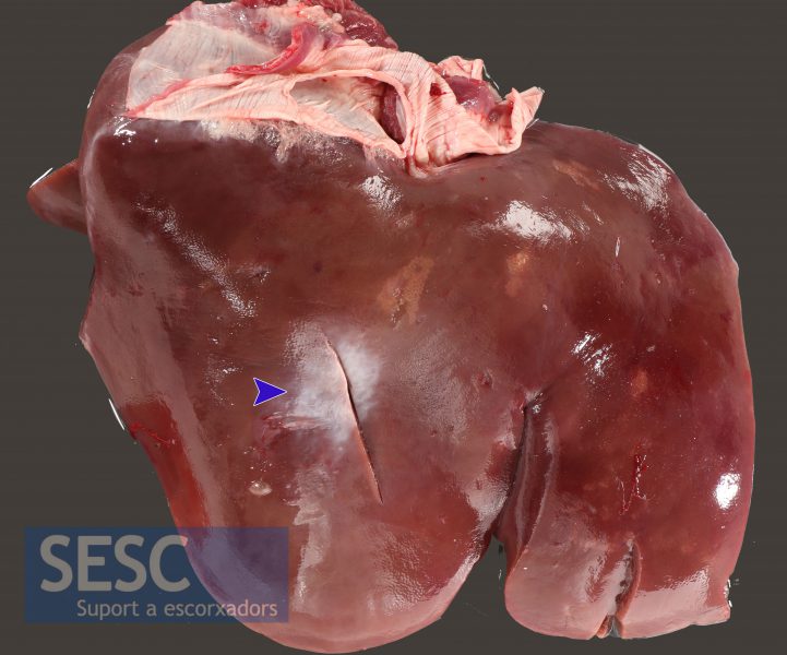
Small ruminant liver that shows a pale area (arrowhead).
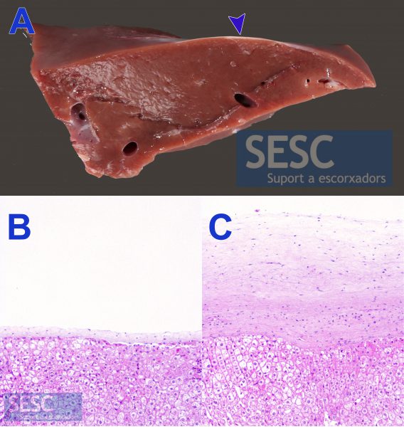
A: When cutting you can see that the alteration affects only the capsule and not the parenchyma. Histology shows that it is a capsule fibrosis area (C), an increase in the thickness of the hepatic capsule, without pathological significance, compared to a normal capsule (B)
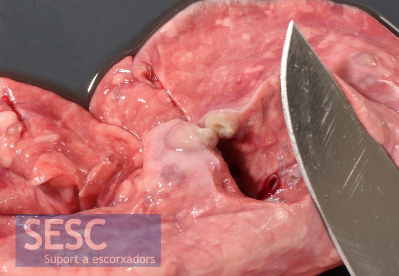
Case 11
Small ruminant liver with an abscess (arrowhead) in the entrance area of the umbilical vein (probably secondary to omfaloflebitis).
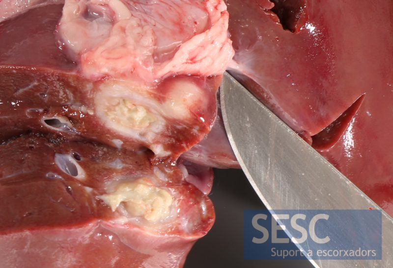
When sectioned we observe the lesion encapsulated with purulent, slightly greenish material, inside.
Necrotic center (left) surrounded by a fibrous capsule (on the right).
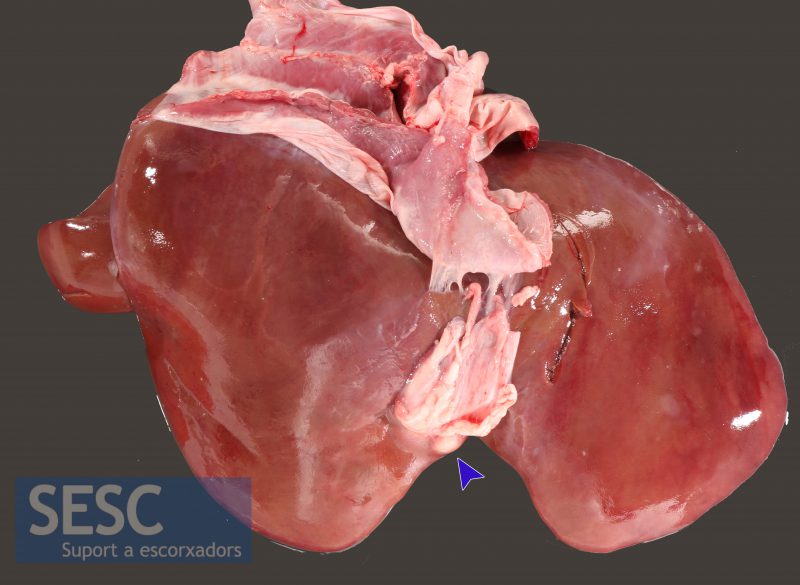
Case 12
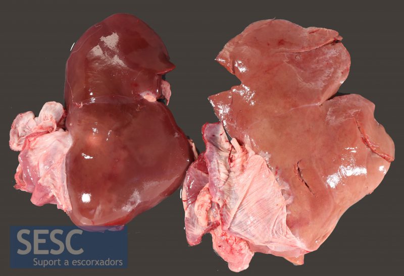
To the right, small ruminant liver with a generalized pale coloring.
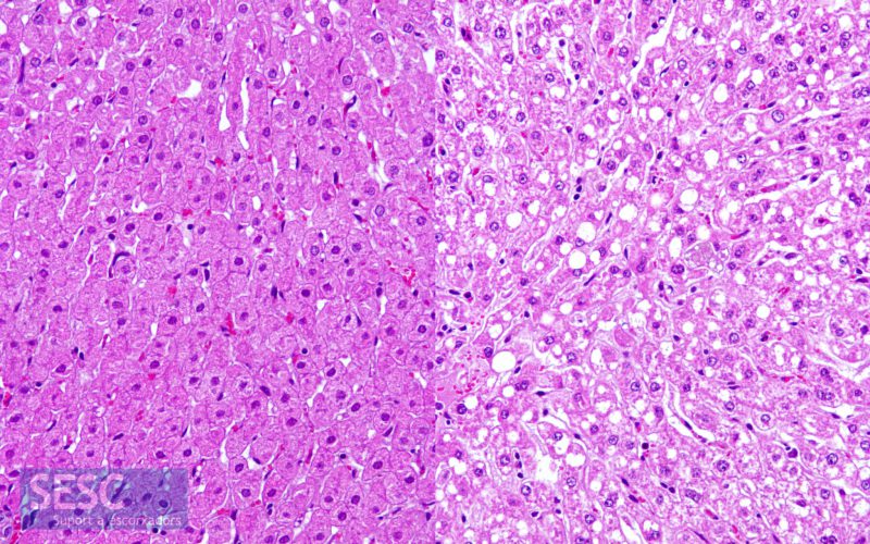
The histopathological study shows that the liver on the right had a hepatic lipidosis (or fat metamorphosis), that is, an accumulation of lipids in vacuoles in the interior of hepatocytes.

1 comment(s)
Bon dia,
Aquest tipus de curs són molt interessants pels SVO. Us demanaria que des del SESC pressioneu, dins les vostres possibilitats, al Departament sobre la necessitat que tots els SVO tinguin accés a aquests cursos.