Black spots in the lungs of a pig
In a 6-month-old, cross-breed pig carcass, multiple small well-defined black nodules were observed, some of them with a whitish center, distributed throughout the whole lung parenchyma. When sectioned, the nodules showed a caseose-like content in the center. On the skin, after scalding, black coloured lesions were seen at the level of the posterior right limb.
The first option, seeing the colour of the lesions, was a metastatic melanoma, but the whitish center that some of the lesions presented did not fit this diagnosis. The histopathological study revealed lesions of multifocal subacute severe embolic-metastatic necrotizing p neumonia with the presence of intralesional bacteria. The black colour was due to areas of haemorrhage surrounding these inflammatory lesions. The whitish caseous center corresponded to necrosis.
The primary origin of the infection is unknown, a possibility is that it was the skin lesion but no skin samples were available for characterization. Another option would be bacterial endocarditis on the atrioventricular valves, in any case the pattern of injury distribution indicates hematogenic dissemination, therefore, it should be considered as a generalized disease.
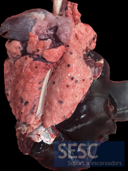
Black nodular lesions of widespread distribution in the pulmonary parenchyma.
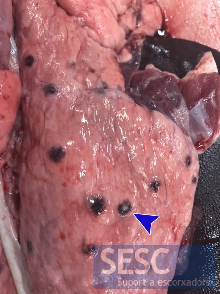
Detail of the lesions, a pale coloured center can be observed in some of them (arrowhead).
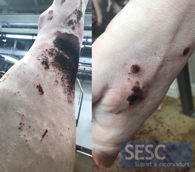
Black skin lesions on the posterior right limb.
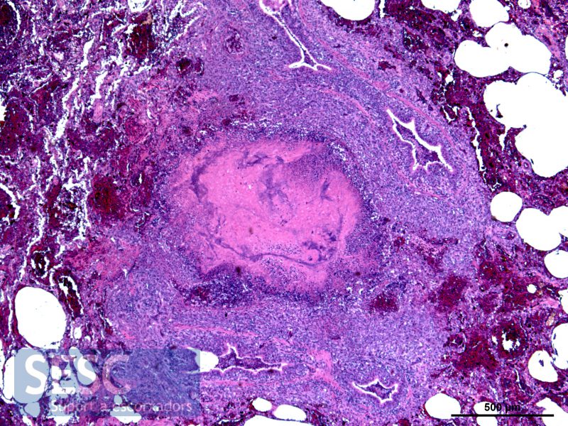
Inflammatory lesion with the presence of bacteria and a necrotic center (lighter pink colour) and surrounded by hemorrhage (reddish colour).
