Bluetongue in Catalonia and its detection in slaughterhouses
As of June 10 of this year 2024, the first focus of blue tongue has been declared in Catalonia since 2009. Since then, cases have been detected in several farms in different counties of the provinces of Barcelona , Girona and Lleida. We refer you to the information published by the Department of Climate Action, Food and Rural Agenda (DAAC) for updated data on the state of the disease in Catalonia. Bluetongue is caused by a virus of the genus Orbivirus, is transmitted by diptera of the genus Culicoides and is not a zoonotic disease.
Bluetongue virus infection can occur in domestic and wild ruminants. It can cause an acute disease in sheep, however it can also present with a generally more moderate clinical condition in cattle and goats depending on the serotype or circulating strain, asymptomatic infections can occur in any species. The serotype 8 is the one circulating in Catalonia and it is manifesting itself clinically in sheep flocks and, to a lesser extent, in cattle.
Finding animals with compatible lesions at the slaughterhouse should not be common. However, in case of suspicion, you can send samples to SESC for diagnosis, with the individual traceability data and the farm of origin. In this post you will find the most relevant clinical and lesional findings, as well as instructions on how to proceed in case of suspected cases.
The most common clinical findings are fever and lethargy, with the animals showing some resistance to movement. It is common to observe edema and/or congestion in the facial area: lips, nose, eyelids, submandibular area... as well as congestion in the coronary arteries. It is possible to observe serous to mucopurulent nasal discharge. When inspecting the oral mucosa, it is possible to observe small hemorrhages or erosions/ulcerations. The tongue can be found relatively increased in size, usually due to edema, and can also present cyanosis.
As for the lesions, apart from those observable in the oral cavity, it is possible to observe other erosions/ulcerations in the gastrointestinal tract, very commonly in the mucosa of the rumen and reticle, where hemorrhages can also be observed. Edema and/or bleeding of the pharyngeal and laryngeal area can be observed, as well as very edematous and congested lungs. Although it is under discussion, the presence of hemorrhages in the pulmonary trunk is described as characteristic bluetongue lesions. In fact, the heart can present myocardial hemorrhages of variable extent, especially apparent in subepicardial and subendocardial locations.
If you have a suspicion, the sample of choice to confirm/dismiss bluetongue is the spleen of the affected animals (or blood with EDTA if there are live animals), from which a PCR will be made to detect the virus. However, we recommend sending other lesioned tissues to SESC in order to reach a diagnosis if blue tongue disease is ruled out. It is also important to consider the differential diagnosis of this disease: the presence of ulcerative lesions in the oral cavity is also potentially compatible with vesicular diseases or other erosive-ulcerative diseases (some diseases are currently absent in Catalonia such as foot-and-mouth disease, vesicular stomatitis and peste des petits ruminants, others of recent outbreak in Catalonia such as epizootic hemorrhagic disease, or other endemic ones such as BVD, among others). The official veterinary services of the slaughterhouse must communicate the suspicions of mandatory declaration diseases to the DACC following the interdepartmental procedure for that purpose, and those identified as category A, and also the blue tongu,e in addition require individualized and urgent notifications.
Below are images of the findings in one of the cases sent to IRTA-CReSA for its necropsy of the first outbreaks detected in Catalonia in 2024. (AC)
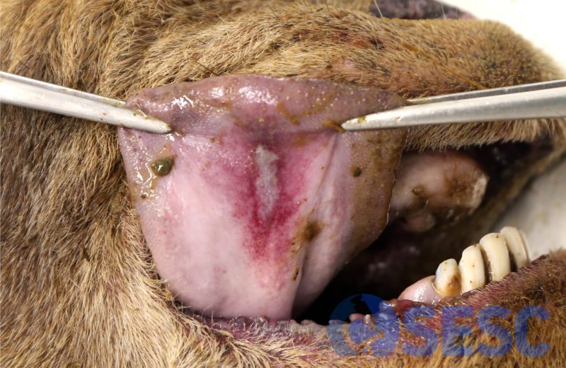
Linear ulcer at the base of the tongue, with a reddened margin.
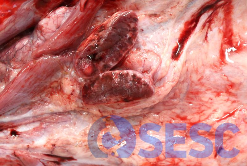
Redness of the submandibular lymph node.
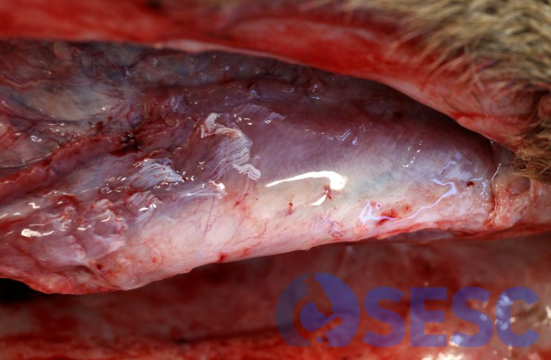
Edema in the region of the submandibular muscles.
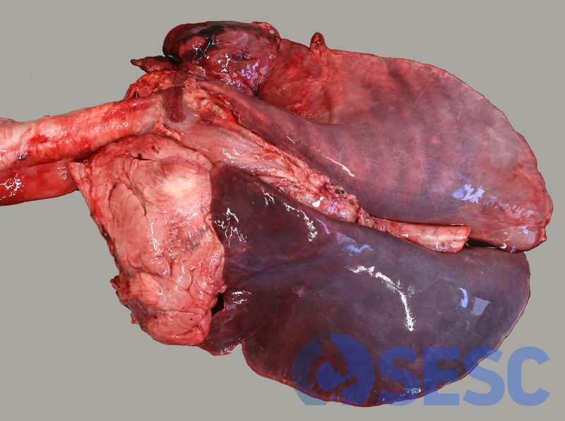
Marked pulmonary edema and congestion.
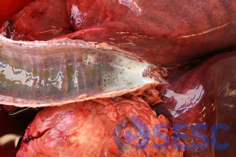
Presence of foam in the trachea, indicative of pulmonary oedema.
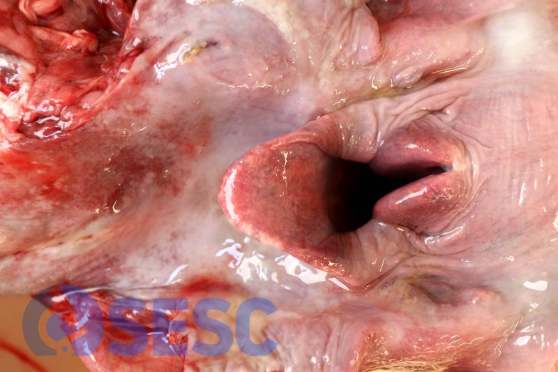
Marked congestion of the larynx.
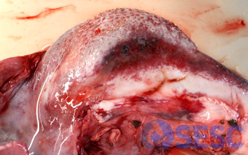
Ulcers and bleeding on the lateral aspect of the tongue.
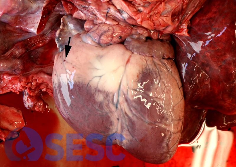
Multiple petechiae in the epicardium. Presence of a small hemorrhage in the serosa of the pulmonary artery (arrow).
