Septicemia due to Staphylococcus spp. in a lamb carcass
Samples were submitted of a 5-month-old ovine carcass from a batch of 28 animals, which presented disseminated lesions throughout the carcass, including liver, lung, spleen, subcutaneous tissue of one limb and lymph nodes. The lesions were small in size, and although it was a lamb, official meat inspectors suspected it could be tuberculosis, hence samples of the different affected organs were submitted to SESC.
Macroscopically all studied viscera presented a generalized multifocal distribution of the lesions, which were of a small size (millimetric, occasionally coalescent), as well as a hard consistency and white color. When sectioned, they revealed a necrotic core which slightly crepitated.
Histologically the lesions corresponded to multifocal areas of lytic necrosis with abundant degenerated neutrophils in their center. The were delimited by a rim of foamy macrophages and a thin band of mature fibrous tissue (early capsule) and variable mononuclear inflammation. Moreover, the necrotic core was frequently mineralized and presented abundant bacterial colonies (cocci) occasionally grouped in tetrads and surrounded by eosinophilic and slightly refringent material (Splendore-Hoeppli material). The morphologic diagnosis was of multifocal to coalescent, severe and chronic pyogranulomatous, necrotizing hepatitis, splenitis, pneumonia and fasciitis, with intralesional bacteria.
These findings were enough to conclude that the lesions were caused by a bacterial septicemia and to rule out tuberculosis. To further define the ethiology of the lesions, a gram stain revealed that the cocci were gram positive. In addition, a microbiological culture of the lesions enabled the isolation of abundant colonies of coagulase positive Staphylococcus in the different samples analysed (liver, hepatic and mediastinic lymph nodes).
The distribution of the lesions (multifocal and generalized) is indicative of a bacterial systemic dissemination. However, the focal localization in the subcutaneous tissue in the posterior left limb suggests that the portal of entry of said bacteria was a cutaneous injury in this region, enabling further dissemination to the viscera. We remind the importance of analysing carcasses in case of gross lesions resembling tuberculosis, in this case the macroscopic appearance of the lesions was not clarifying due to their small size.(AC)
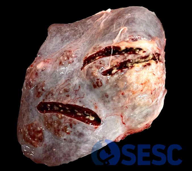
Lamb spleen that presented white, multifocal nodular millimetric lesions, mainly apparent when the viscera was sectioned.
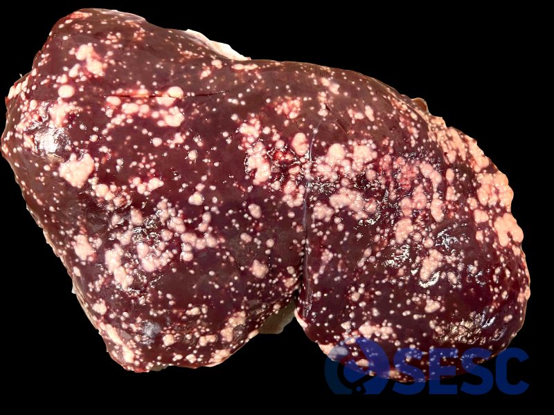
Lamb liver presenting the same lesions with a multifocal miliary generalized distribution, prominent through the hepatic capsule.
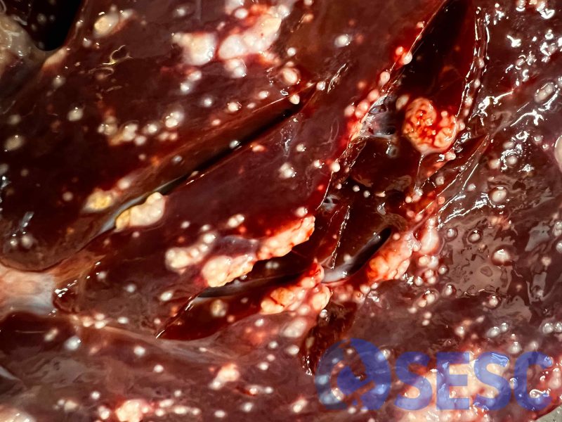
Detail of the same liver, revealing the appearance of the lesions. They present a very small size, although they often coalesce and create larger lesions. Also, the cut section reveals lesions within the parenchyma and the necrotic/purulent content within the lesions.
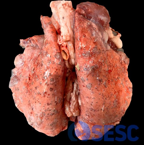
Lamb lung which presents similar lesions, with a multifocal generalized distribution (pattern of embolic pneumonia or bacterial systemic dissemination).
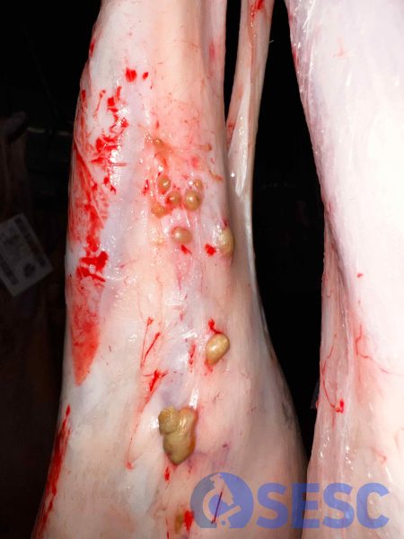
Lesions in the left posterior limb, between the subcutaneous tissue and the muscular fascia. Histologically they are identical to the rest of the lesions. Their presence in this only localization within the subcutaneous tissue suggests that a cutaneous lesion in this region was the portal of entry.
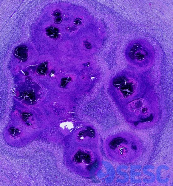
Histology of some of the lesions, which shows coalescence of the lesions upon contact of the necrotic cores.
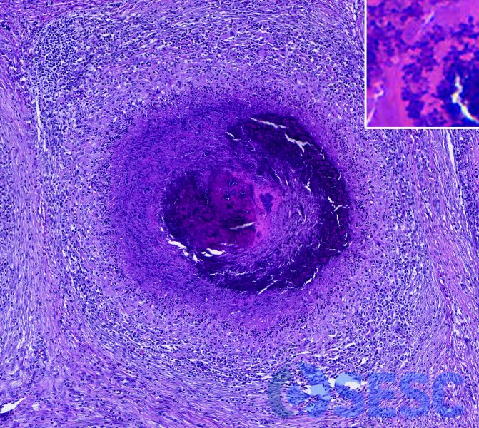
One of the lesions, in which the mineralized core can be seen within the necrotic debris and degenerated neutrophils. It is demarcated by foamy macrophages, fibrous tissue, and mononuclear infiltrate. At higher magnification (inset) abundant cocci are observed, surrounded by intense eosinophil material (Splendore-Hoeppli).
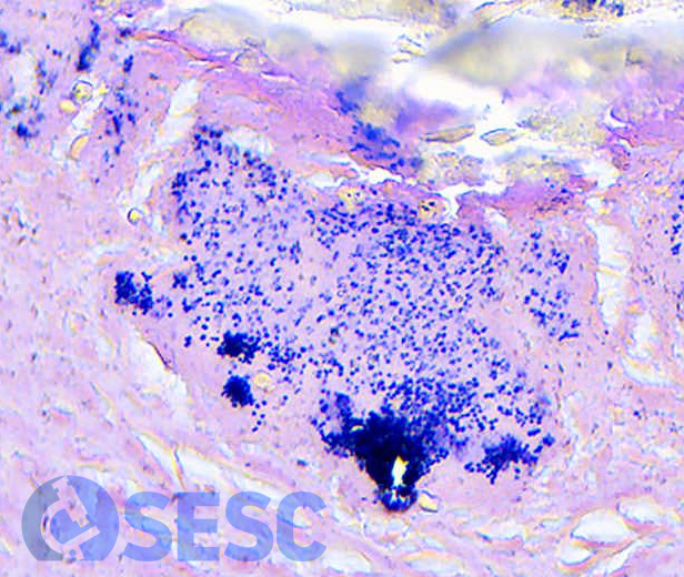
Gram stain reveals that the cocci are Gram positive, consistent with the identification of Staphylococccus spp.
