Bovine carcass with a “gritty” subcutaneous appearance
Histopathology revealed the presence of multiple protozoan intracellular cysts among the muscular fibres and subcutanous adipous tissue cells. The morphology of the cysts was typical of the Sarcocystidae protozoan family, very probably from the genus Besnoitia, in cattle the most common species is B.besnoiti.

Carcass with a congestive appearance and the presence of ecchymosis.

Detail of the altered texture of the subcutaneous adipous tissue. Note the presence of minuscule whitish nodules of gritty appearance, which correspond to the Besnoitia bradyzoites cysts, big enough to be seen with the naked eye.
, of a considerable size, containing numerous besnoitia bradyzoites among the muscle fibers (right) and associated to an inflammatory reaction (top, left).")
Haematoxylin eosin microphotography. One of the multiple cysts observed (center), of a considerable size, containing numerous besnoitia bradyzoites among the muscle fibers (right) and associated to an inflammatory reaction (top, left).
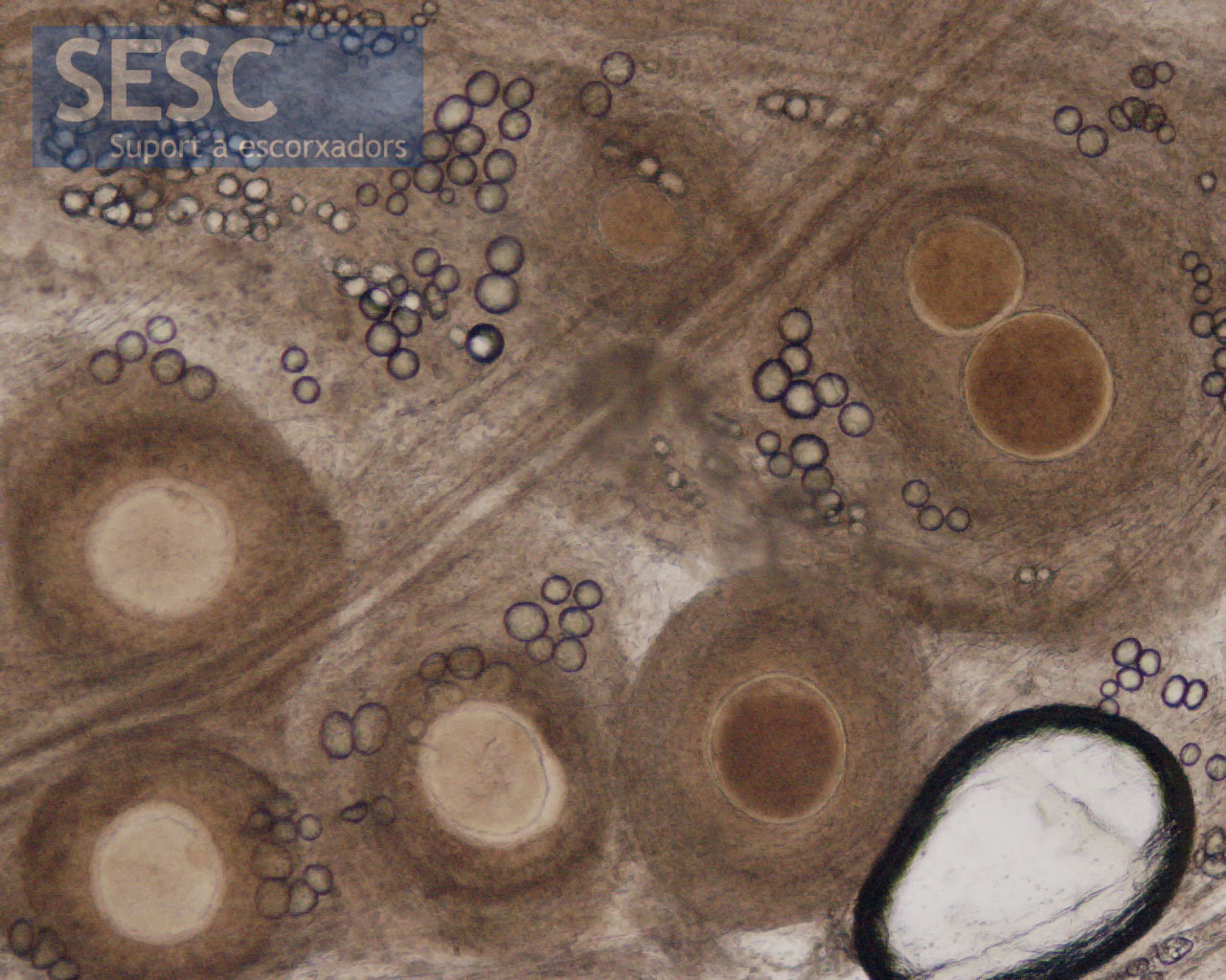
Direct microscopic observation of connective tissue of an animal with besnoitiasis. The spherical structures are the cysts.
