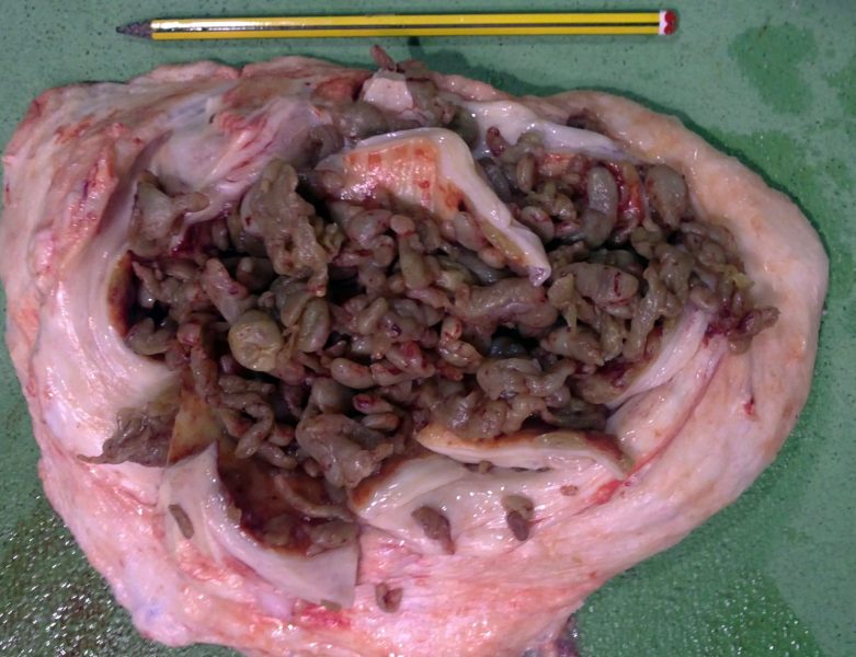
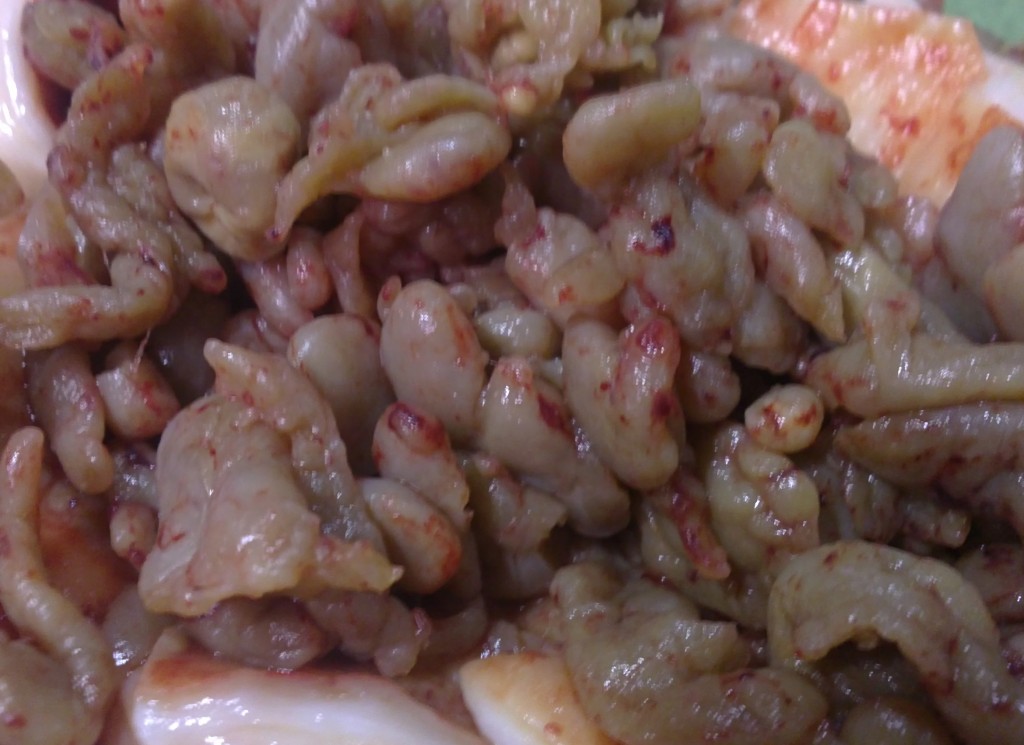
Subcutaneous nodule in the flank of a 3 years old friesian cow
When sectioned, an internal cavity was disclosed filled up with amorphous structures, of smooth surface, gelatinous consistency, brownish colouration and of the size of a bean or bigger. These structures did not adhere to the fibrous capsule or between themselves.
Histopathology revealed an abundant amount of granulation tissue, chronic in nature, constituted by well organized, well vascularised, dense collagen fascicles. This granulation tissue progressed towards the center, being more mature at the periphery of the lesion.
In the core of the cyst the tissue is denatured (hyalinized) and with a necrotic appearance. The structures within the cyst show no histological structures consisting basically of necrotic material.
It most probably is an exhuberant and chronic healing reaction. A possible origin of which could be the injection of a subcutaneous substance.


