Brown fat horse carcass
In a 5 years old mare a discoloration of adipose tissue was observed. It presented a brownish coloration of both subcutaneous and visceral fat (see Figures 1 and 2).
The inspectors differential was of a xanthomatosis. In veterinary medicine this term refers to a type of degeneration of muscle fibers, also called lipofuscinosis, which is characterised by a lipofuscin pigment deposition in muscle cells that gives a brownish-grayish color. In human medicine, however, xanthomatosis refers to the occurrence of xanthomas or adipose tissue (nodular) deposits enriched in cholesterol and foam cells. The latter presentation is also described in birds.
In the present case, histologically, pigment deposits were observed in the adipose tissue in the form of spherical structures of various colors and sizes (see Figures 3, 4 and 5) amongst normal adipocytes. These structures were most likely within phagocytic cells, but it could not be clearly discerned. Fat necrosis or presence of inflammatory lesions were not obserevd. The conjunctive tissue had small basophilic coloured areas with calcium deposits and some swelling. Muscle cells showed no alterations.
The microscopic appearance of the lesion is suggestive of a wear-and-tear pigment, such as lipofuscin or ceroid, which could be a consequence of adipose tissue degeneration. In this sense the lesion could be classified as a lipodystrophy.
In an attempt to characterize the lesion PAS staining of the cells was performed but it did not stain the droplets (lipofuscin is normally PAS +).
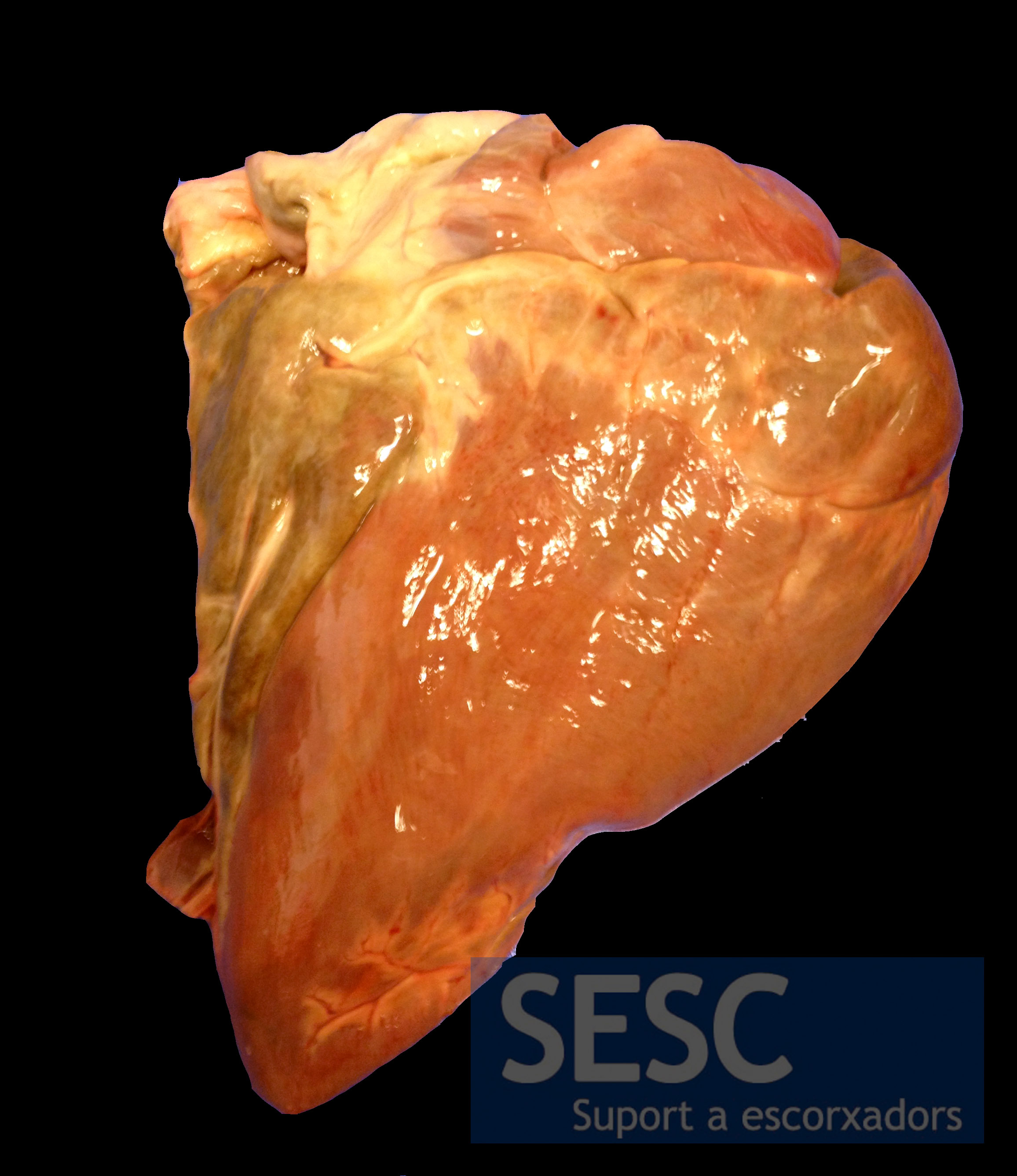
Adipose tissue showed brown discoloration. In the picture this is evident in the epicardial fat.
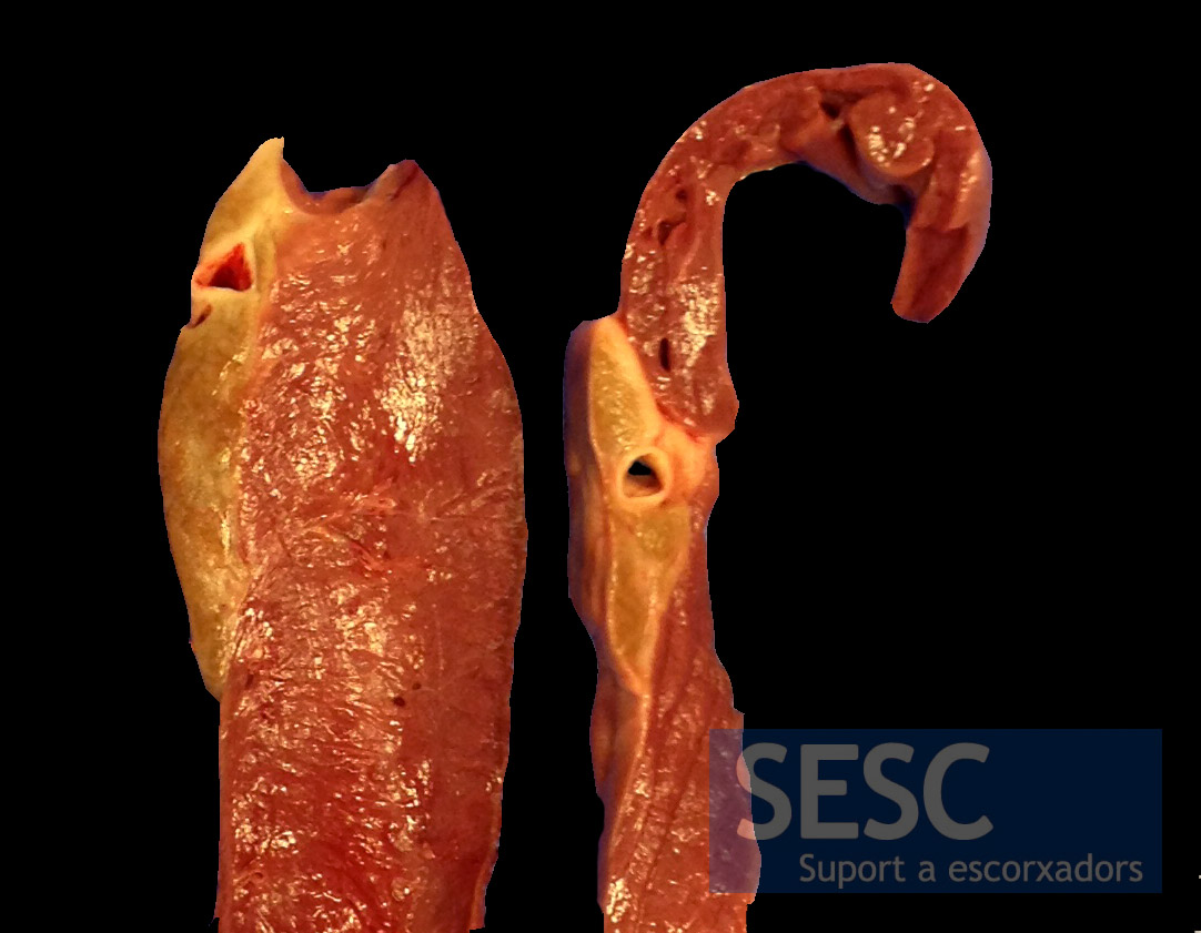
When sectioned it can be seen that the coloring affects the entire thickness of adipose tissue.
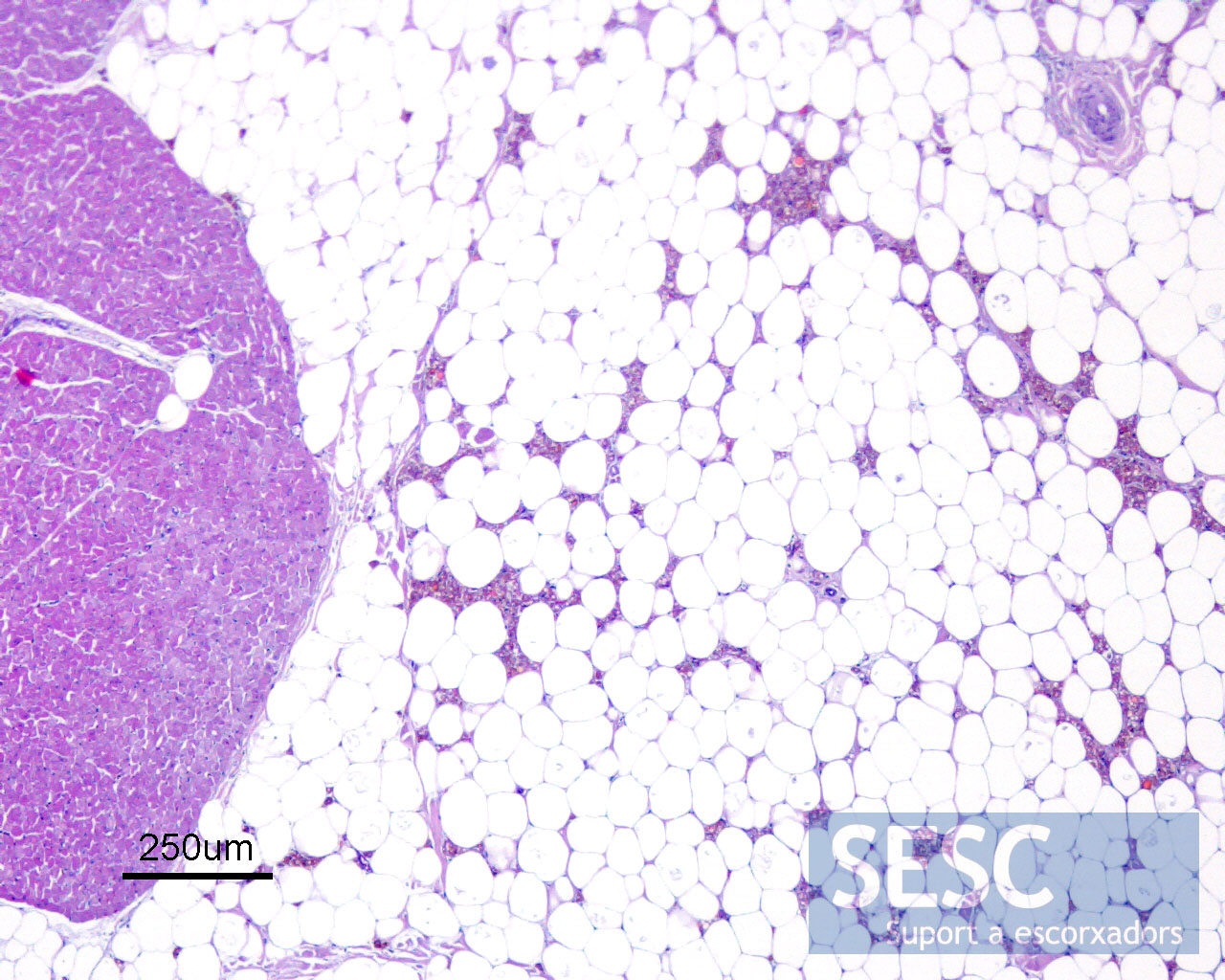
Pigment deposits could be observed between normal cells of the adipose tissue.

Microscopically this image was oberved: clumps of pigment, probably within phagocytic cells, amongst normal-appearing adipocytes.
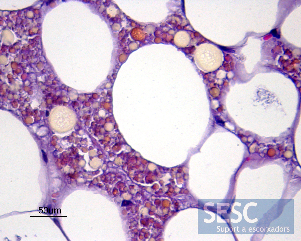
No fat necrosis or inflammation were observed. The droplets had various colorations such as brown, ocher and orange.
