Granulomatous myositis and steatitis in a mare
In a 15-year-old mare of the Spanish-Breton breed, multiple nodular lesions, small (<2mm) and of generalized miliary distribution were observed in the musculature and adipose tissue of the carcass.
Histopathological examination evidenced, in both tissues, granulomatous myositis and setatitis lesions with abundant eosinophilic polymorphonuclear leukocytes. This type of lesion is compatible with a parasitic etiology.
In fact, some muscle fibers were spotted with Sarcocystis spp. cysts within their cytoplasm, but those were not associated with the lesions.
PAS, Ziehl Neelsen, Grocott and Gram staining failed to demonstrate the presence of other possible agents such as fungi, bacteria, mycobacteria or other parasites.
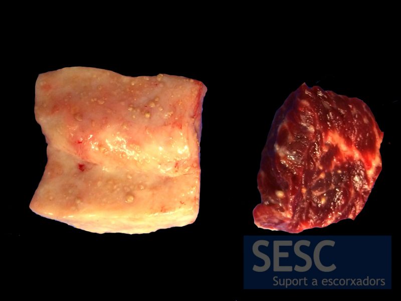
Whitish nodular lesions in the fat (left) and muscle tissue (right)
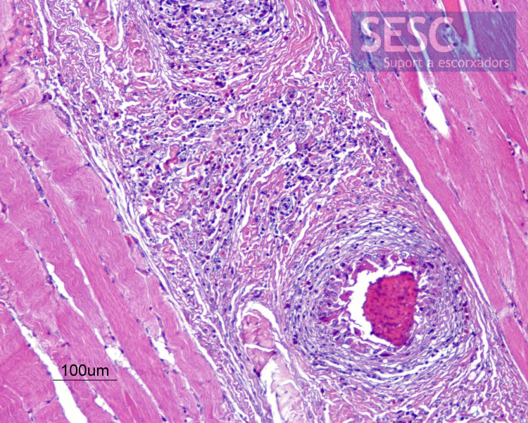
Granulomatous myositis.
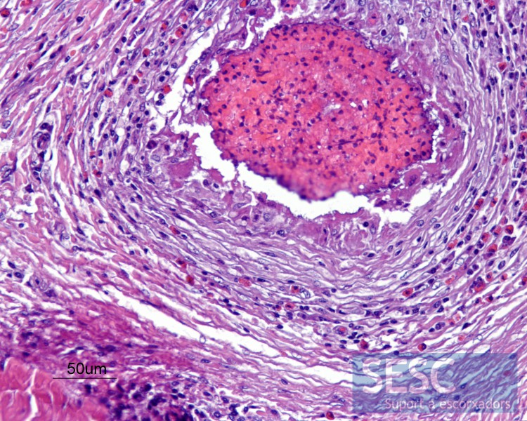
Granulomatous myositis. Central necrotic area surrounded by fibrosis and inflammatory infiltrate rich in eosinophilic polymorphonuclear leukocytes.
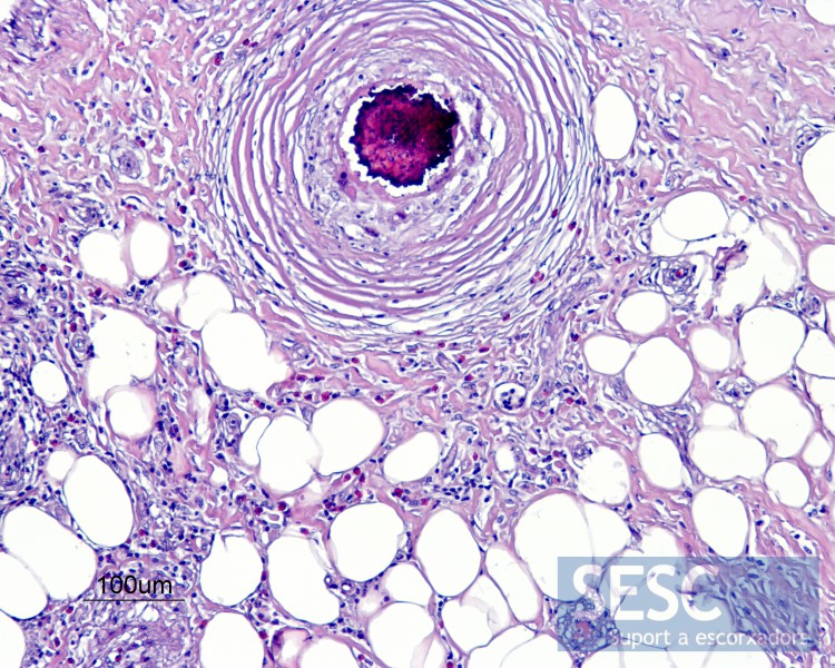
Granulomatous Steatitis. The same type of lesion observed in the muscle could be observed in the adipose tissue.
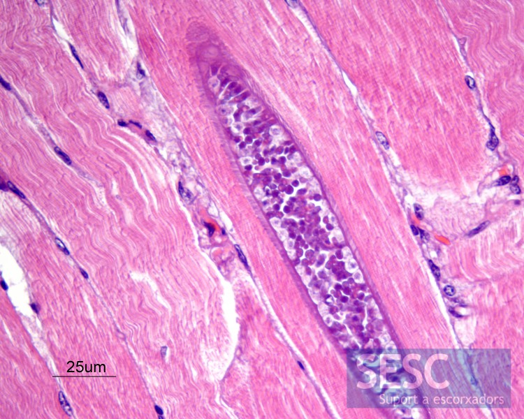
Sarcocystis spp. cyst within a muscle fiber..

1 comment(s)
Parasitic aetiology, not Sarcocystis. Granulomas contain degenerate parasites no longer recognizable. Maybe Taenia sp or aberrant migrating nematode larvae.