Horse carcasses with yellow fat disease
In the last months 4 cases of horse carcasses with an abnormal yellowish-brown coloration of adipose tissue have been submitted.
All cases consisted of yellow fat disease cases which is a chronic granulomatous steatitis (it would fall within a more general concept of Lipodystrophy, referring to an alteration of adipose tissue). We have previously described this condition in cattle.
Briefly, this disease consists in the fact that due to a lack of reducing power lipids are oxidized and what we observe in the microscope (which is what gives the yellowish-brown color to the fat) is the accumulation of pigments resulting from the degradation of these lipids, particularly within macrophages. This oxidation of lipids causes a macrophagic inflammatory reaction (=diffuse granulomatous). These pigments are known by the name of ceroid, which is a type of lipofuscin.
Earlier, this cases were diagnosed with the term xanthomatosis, but it is a term in disuse (no longer appears in recent textbooks) and therefore we recommend to stop using it.
Regarding public health, since it is a generalized inflammation of adipose tissue, we consider the carcasses unfit for consumption, but essentially no sanitary risk exists other than the organoleptic alteration of both color and, probably, taste (oxidized, rancid fat).
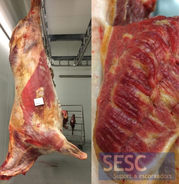
Change in coloration of adipose tissue. On the right detail of a section of muscle.
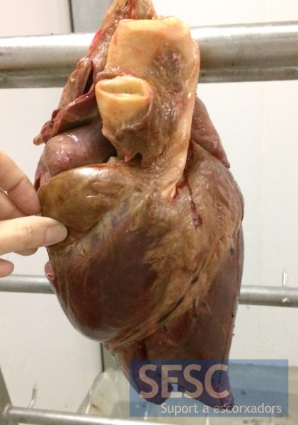
The adipose tissue of the epicardium also shows an evident color change.
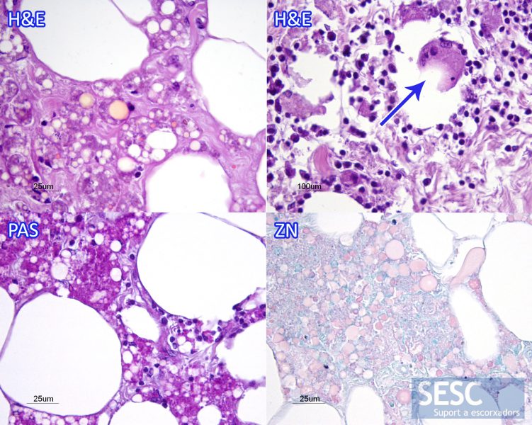
Anatomopathological study. The preparations stained with hematoxylin and eosin (H&E) allow the observation of the presence of macrophages in adipose tissue with intracytoplasmic pigment accumulation in the form of small orange spheres. In some sections there was also an overt inflammation of adipose tissue with presence of multinucleated giant cells (arrow). In this case the pigment was stained intensely with the Periodic Acid Shiff (PAS) stain that labels polysaccharides. The Ziehl Neelsen staining (ZN) stained with variable intensity the pigment accumulations.
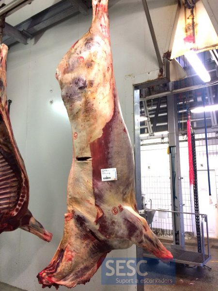
Another example of yellow fat disease.
