BLOG
SESC case archive


16/11/2008 | Poultry
0

Inclusion body hepatitis

When the coelomic cavity was opened ascites and hepatomegaly were observed accompanied by paleness of the liver, which was yellow colored and friable.

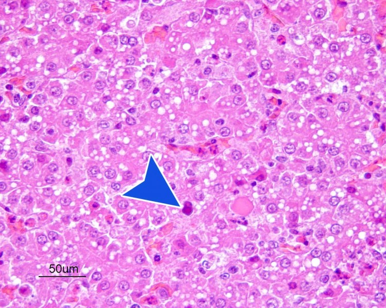
Pathological examination revealed the presence of inclusion body hepatitis, caused by a Type I avian adenovirus.

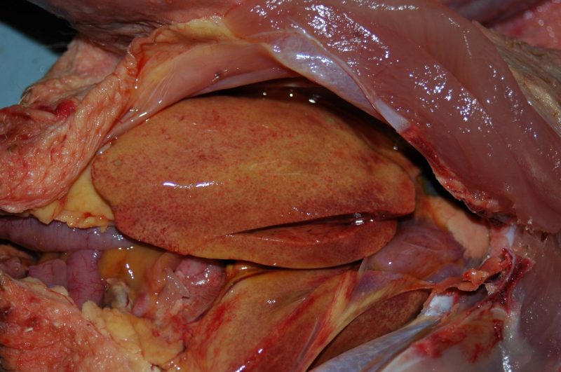
Yellowish liver with petechiae and hepatomegaly.

Yellowish liver with petechiae and hepatomegaly.

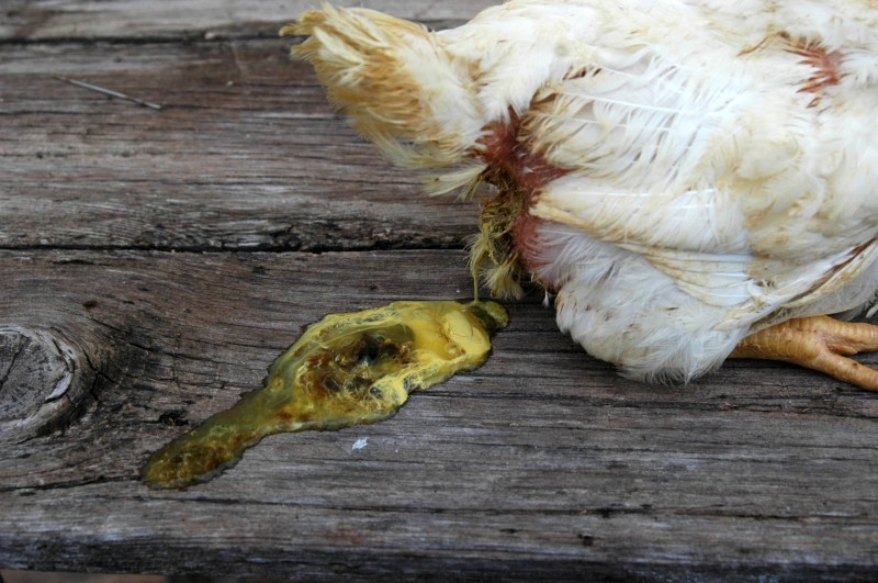
Feces of liquid consistency and altered color.

Feces of liquid consistency and altered color.

Hepatocyte with eosinophilic intranuclear inclusion body characteristic of this disease.

Hepatocyte with eosinophilic intranuclear inclusion body characteristic of this disease.


Add Comment

Your email address will not be published. Required fields are marked *