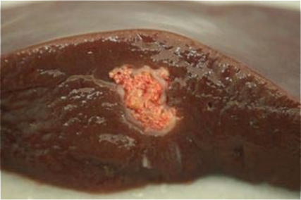
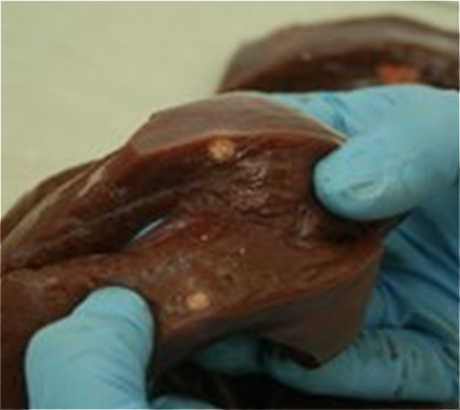
Hydatidosis (E.granulosus).
The presence of nodular lesions in the liver was observed in two different calf groups, in each case there were three affected animals. The differential diagnosis proposed by inspectors included Tuberculosis.
Histopathological examination revealed necrotizing granulomatous-like lesions with presence of abundant eosinophilic polymorphonuclear leukocytes. This type of lesion is associated with hydatid cysts in different stages of involution and degeneration as a result of the host inflammatory reaction.
Hydatid cysts are the the form taken in the intermediate host by Echinococcus granulosus, the adult form of which is an intestinal tapeworm whose definitive host are canids.
The oncospheres hatch when ingested by intermediate hosts (ruminants, pigs or people, etc.) penetrate the intestine and usually form hydatid cysts in the liver, or if they reach the cava vein though lymph vessels, can occur frequently in the lungs, or in any other organ.
Cystic lesions may contain fluid inside with multiple protoescolices (hydatid sand) and are usually associated with a granulomatous reaction. The size can vary from a few millimeters up to 10 cm in diameter.
It is a zoonosis.
In this case, we present degenerate cysts associated lesions and necrotic (often mineralized) that may be confused with tuberculous granulomas.

Intraparenchymal, encapsulated, white nodule in the liver.

Smaller size, irregular, whitish, intraparenchymal nodule in the liver.

