Lymphoid leukemia in broilers due to Marek’s virus infection
The veterinary students at the Universitat Autònoma de Barcelona (UAB) perform necropsy practices with avian carcasses, usually with broilers that arrive dead at the slaughterhouse. In one of the latter sessions, severe lesions were found consistently in 12 broilers that apparently came from the same batch. Samples were submitted to SESC to investigate this case.
The macroscopic examination conducted by the students found as the main alteration a very marked splenomegaly, which was enlarged by 3-4 times the normal size. As a comparison, the spleens were larger than the hart and -in the most severe cases- almost as big as the gizzard. Also, they presented a soft consistency and when sectioned, the parenchyma bulged over the capsule. In some animals, white multifocal areas were also seen in the liver and/or kidneys. In contrast, no macroscopic alterations were observed in the sciatic nerves. Samples of spleen, liver and kidney from 6 affected animals were formalin fixed, and fresh liver and spleen samples were collected.
Under histopathological evaluation, a marked proliferation of round cells was observed, which occupied a great proportion of the evaluated spleens, as well as in the white foci of the liver and kidneys. The cells morphologically resembled lymphocytes but with a larger nucleus. Also, interestingly, the cells in the liver were predominantly located within hepatic sinusoids. Through immunohistochemical assays against CD3 and CD20, the cells are confirmed to be of T lymphocytic lineage.
The majority of the proliferations of T cells in poultry are caused by the Marek Disease virus (MDV), so we submitted fresh spleen samples to perform molecular techniques. PCRs were performed against avian leucosis virus and reticuloentotheliosis virus, which resulted negative. In contrast, a PCR against MDV resulted in a high viral load. A PCR to discriminate vaccinal virus with field strains was performed, which confirmed that the virus was a field strain. Finally, sequencing of the strain was performed, which permitted to classify it as a very virulent plus pathotype (vv+MDV).
The Marek disease observed in these cases doesn’t exactly match with any of the classical forms (visceral, neural, flaccid paralysis…). In contrast, it coincides with the Marek lymphocytic leukemia form which has been very rarely described, associated to very virulent strains. This form is differentiated from the classical lymphoma by means of macroscopic evaluation (diffuse splenomegaly) and histologic evaluation (presence of neoplastic cells within blood vessels rather than in solid growths). As this is such a rare presentation, there is no explanation as to why in this batch there are so many animals with this presentation. (AC)
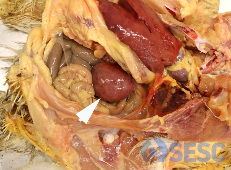
Very marked splenomegaly.
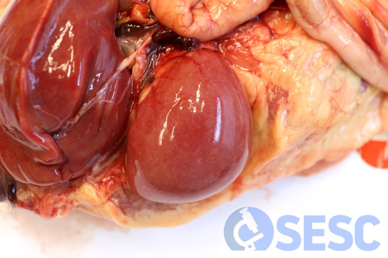
Very marked splenomegaly. The spleen presents a pale coloration, with a slightly heterogeneous appearance (generalized white spots).
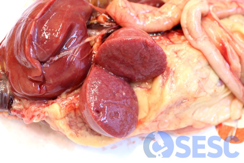
The same spleen when sectioned. The parenchyma bulges over the capsule, indicative of the cellular proliferation that is expanding it.
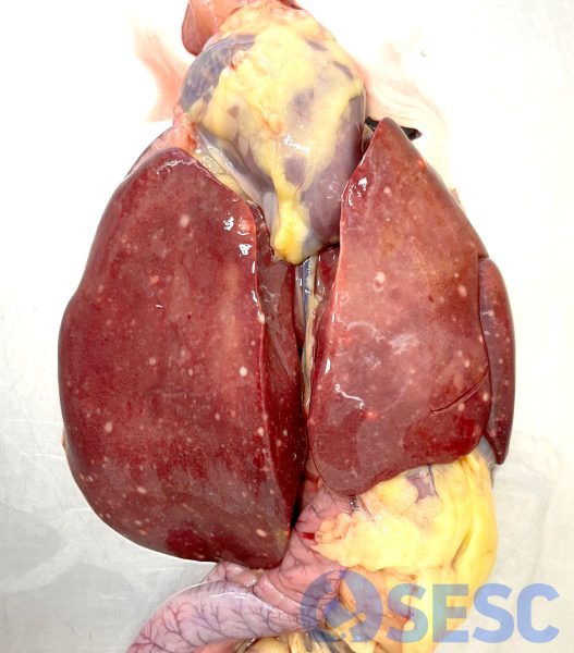
Liver of one of the affected animals, which presents multifocal white areas in a generalized distribution.
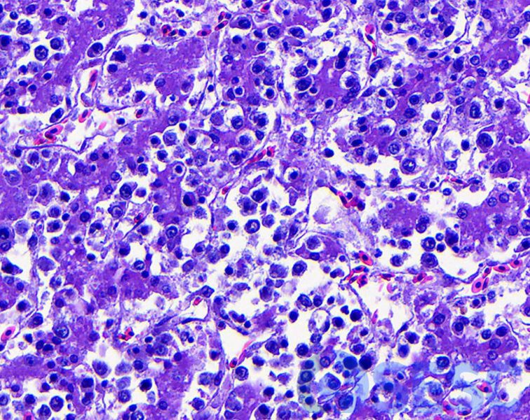
Histology of the liver. Abundant lymphocytes of large size are expanding hepatic sinusoids.
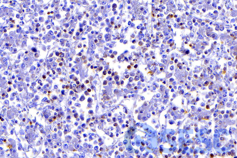
The immunohistochemical technique against CD3 confirms that the neoplastic cells are T lymphocytes.
