Arthritis due to Staphylococcus aureus in quails
An inquiry was submitted regarding different batches of 5-week-old quails which came from the same farm. They arrived at different days but some of them shared a common alteration: unilateral enlargement at the tarsal region. The official meat inspectors suspected of a nutritional or infectious process, so they submitted samples of 8 affected limbs at SESC.
Upon receiving the samples, a very pronounced enlargement of the tarsal region was noted in the area corresponding to the tibiotarsus-tarsusmetatarsus joint. When sectioned, it corresponded to the accumulation of caseous material within the articular capsule and the gastrocnemius’ tendon shaft. When the histopathological study of the samples was performed, this material corresponded to necrotic debris, heterophils and fibrin, confirming that it consisted of fibrino-suppurative arthritis. Additionally, bacterial colonies were observed, showing coccoid morphology and aggrupation in clusters. A gram stain was performed, which revealed gram-positive cocci.
Samples of the joints were submitted for microbiological culture, which yielded pure and abundant growth of Staphylococcus aureus, which coincides with the morphology and gram-positivity of the observed bacteria. The presence of arthritis due to S. aureus in poultry is often the consequence of entry of this bacterium secondary to skin lesions, mucosal inflammation, omphalitis, parenteral vaccinations, etc. and its posterior hematologic dissemination. (AC).
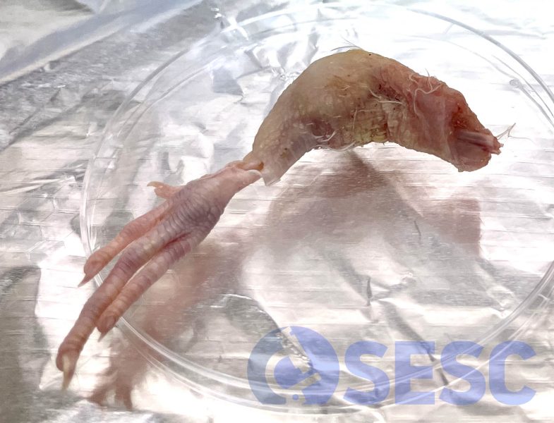
Quail’s posterior limb. A very marked enlargement of the limb can be observed in the region of the tibiotarsus-tarsometatarsus joint.
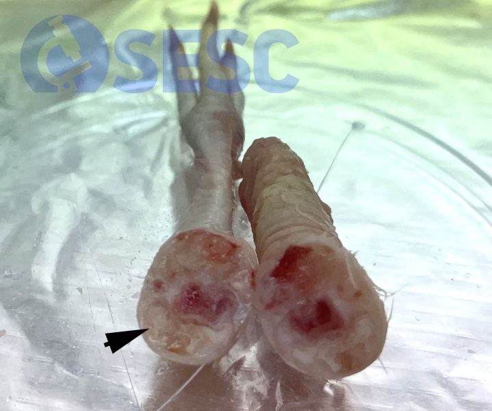
Quail’s posterior limb, sectioned. The enlargement of the limb corresponds to the accumulation of caseous material within the articular capsule (arrow).
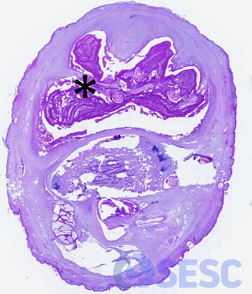
Histological section of the limb, at low magnification, which shows a marked distension of the articular capsule due to the accumulation of exudate (asterisk), which surrounds the gastrocnemius tendon.
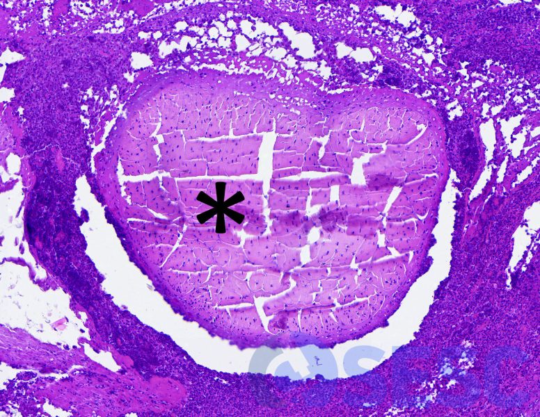
At higher magnification, the exudate surrounds the gastrocnemius tendon (asterisk), composed of viable and degenerated heterophils, necrotic debris and fibrin.
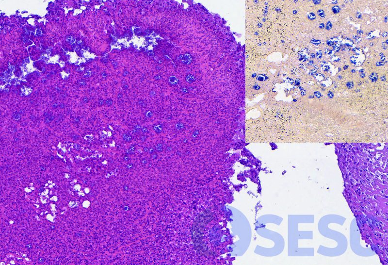
Histological section at the margin of the lesion, where the exudate appears near the articular capsule. Within the necrotic debris, abundant bacterial colonies can be observed grouped in clusters. These colonies appear gram-positive under a gram stain (insert).
