Splenomegaly and peritoneal lesions in a pig carcass
The histopathological study showed, both in the spleen and in the peritoneal lesions, a neoplastic round cell proliferation surrounded by an abundant proliferation of connective tissue. Polymorphonuclear eosinophilic leukocytes could be observed infiltrating this tissue. Morphological diagnosis was compatible with a Mastocytoma.
To confirm the diagnosis immunohistochemistry against C-kit receptor was done, with a positive outcome. Toluidin Blue stain revealed the presence of intracytoplasmic granules in the neoplastic mast cells.
It is a rare neoplasm in pigs, we had previously described a case affecting the skin: Cutaneous Mastocytoma.
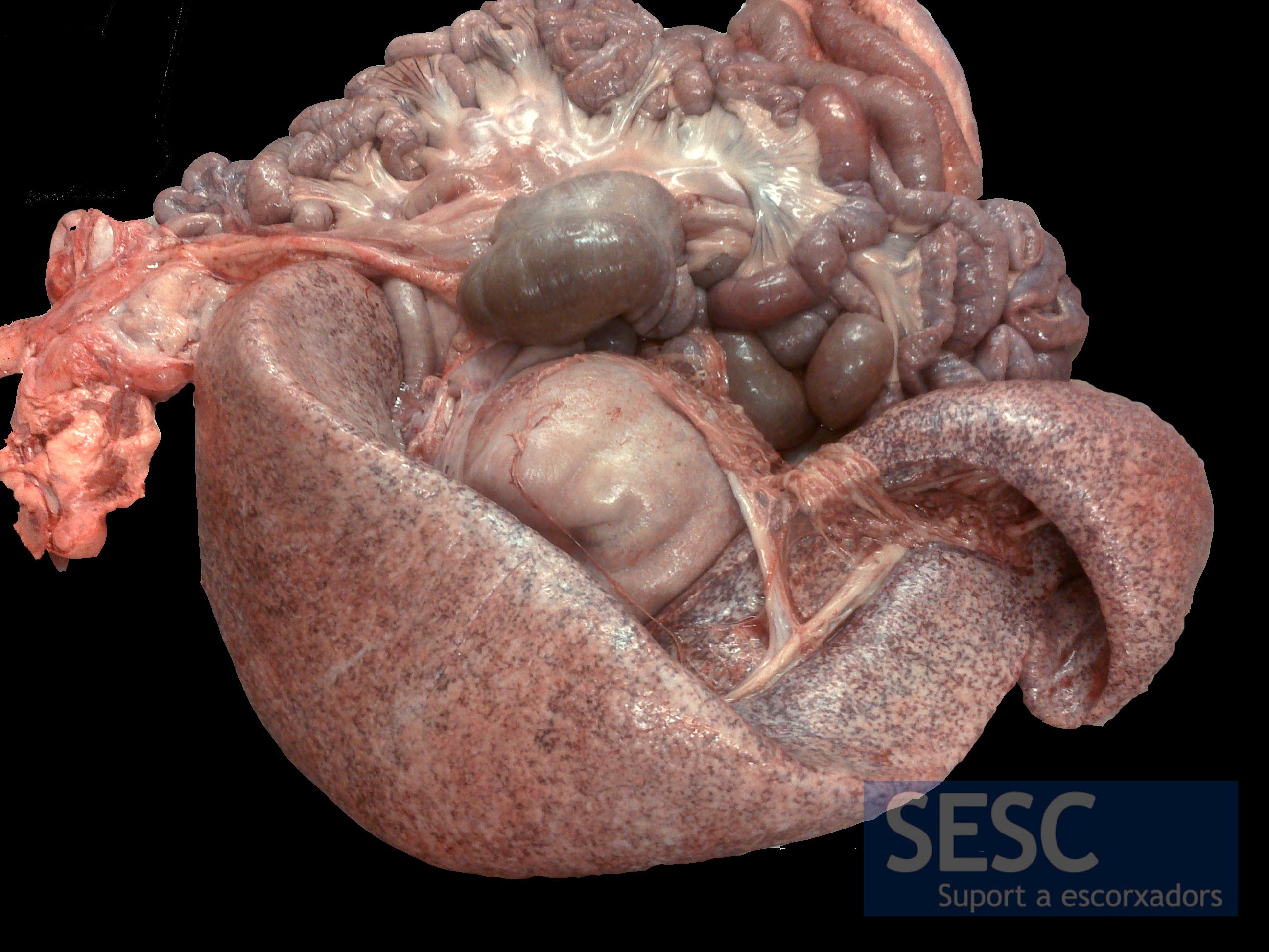
Marked increase in size of the spleen, which presents a pale appearance.
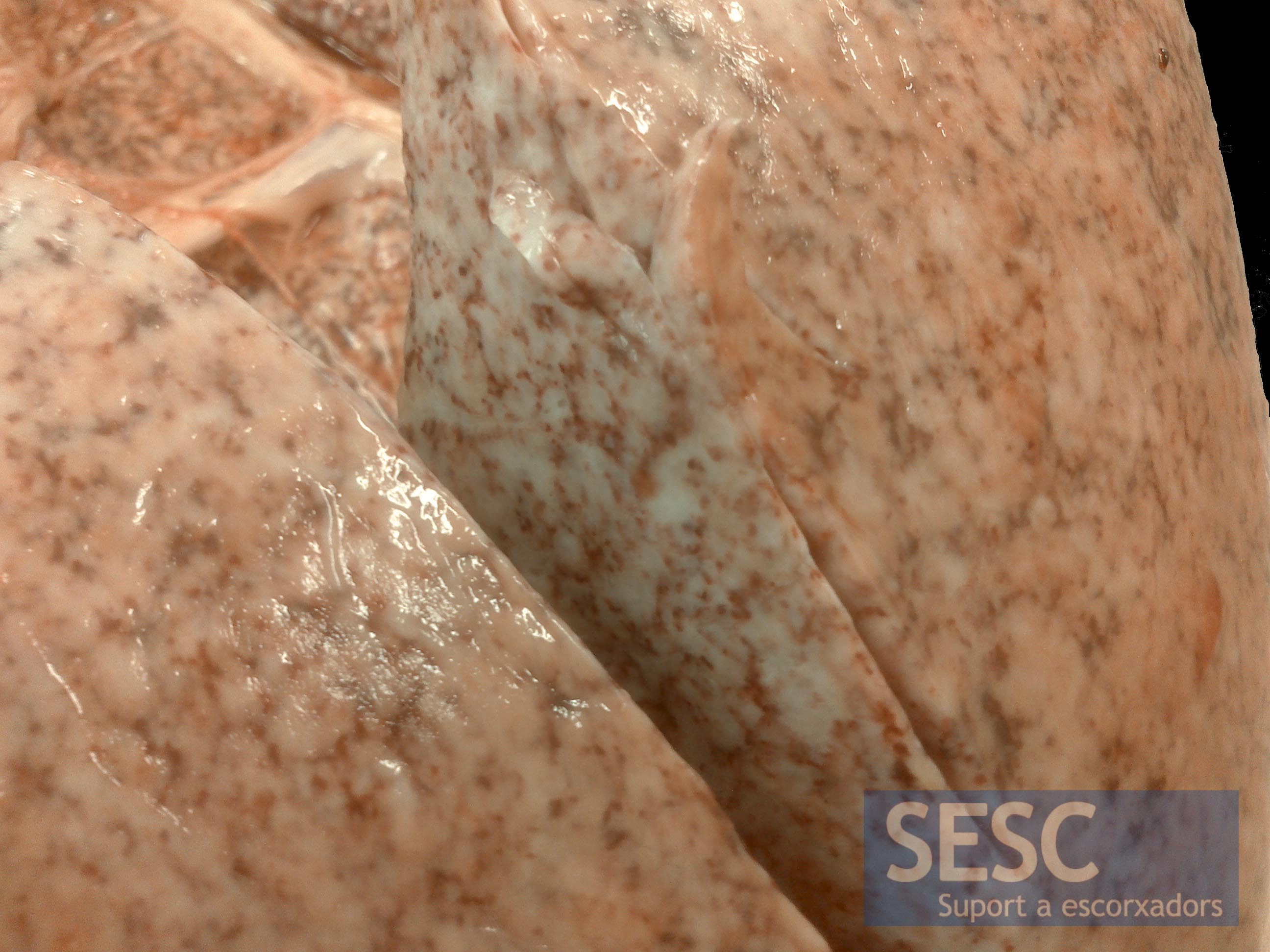
Detail of the marble-like appearance of the splenic parenchyma.
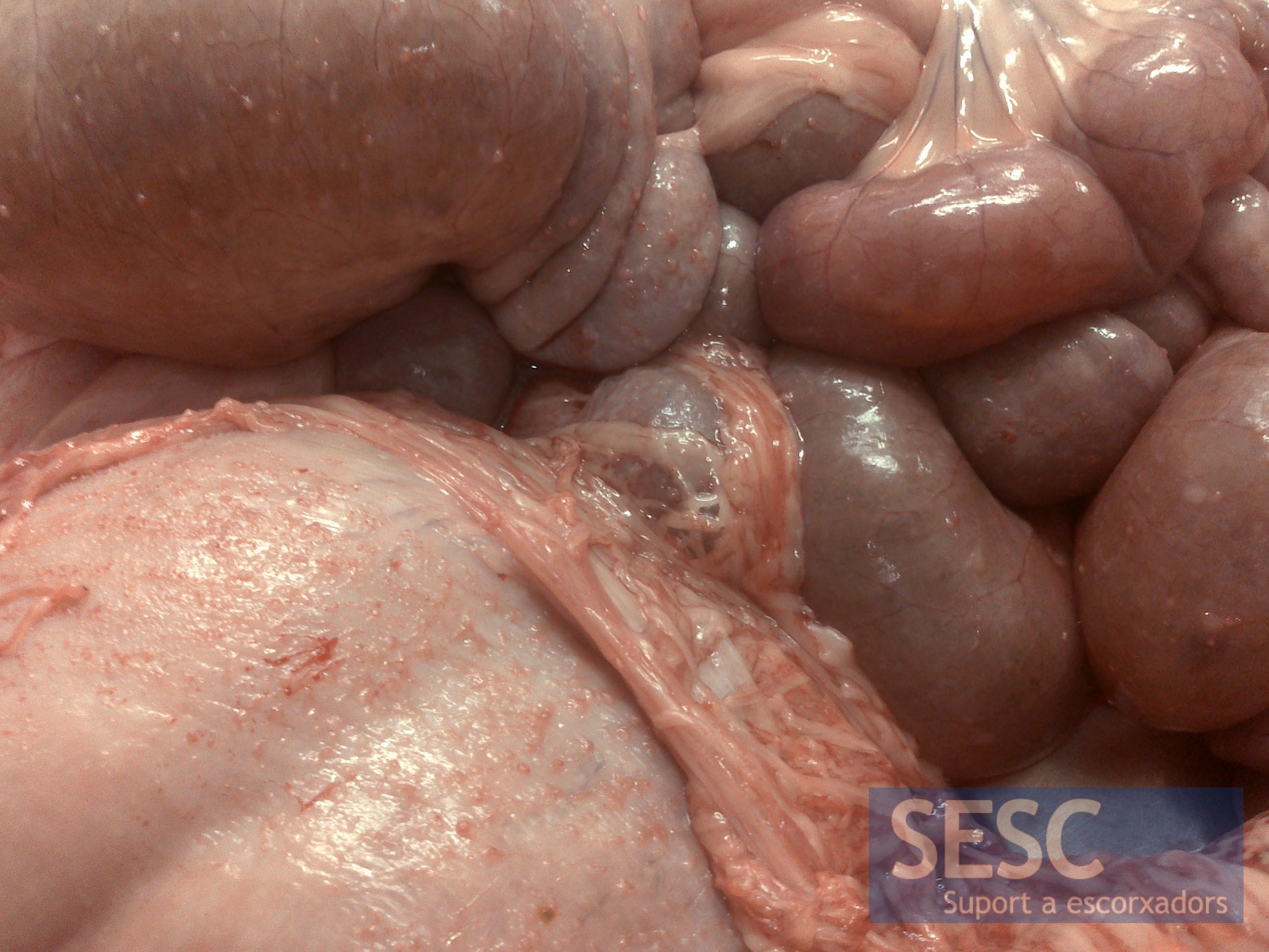
Miliary nodular lesions in the serous membranes of the abdominal cavity (intestines, stomach, etc.)
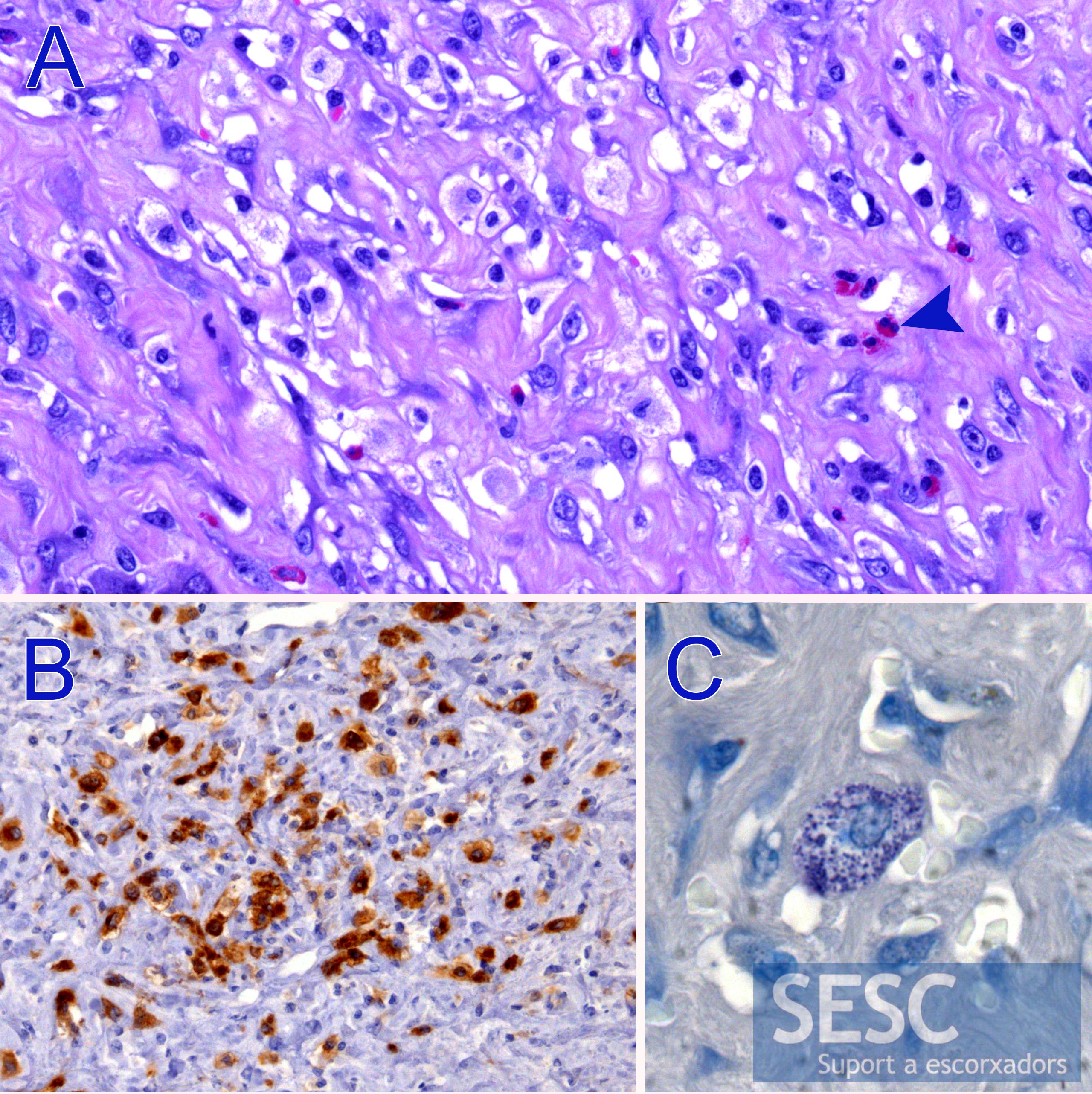
Histopathological study. A: H&E staining shows neoplastic mast cells surrounded by a fibrous matrix and presence of eosinophils infiltrating the mass (arrowhead). B: These mast cells were positive for the immunohistochemical marker against C-kit receptor. C: Toluidine blue staining showed the presence of intracytoplasmic granules.

1 comment(s)
Comments from Veterinary Pathology groups in LinkedIn:
by LuAnn mckinney:
Nice case! Thanks for sharing.
by Ana Rosa De Sousa Resendes:
beautiful case! thanks for sharing
by Norman Barlow:
That was a surprise diagnosis in a 6-month old pig.