Granulomatous pneumonia in a lamb
Histopathology showed multiple nodular necrotizing-granulomatous lesions partially mineralized and with the typical concentric arrangement of caseous lymphadenitis (caused by C.pseudotuberculosis , as evidenced by the positive result in the Gram stain).
However microbiological culture yielded solely a pure culture of Pasteruella multocida . It might be a coinfection, but the granulomatous lesions described are not typical of Pasteurella .
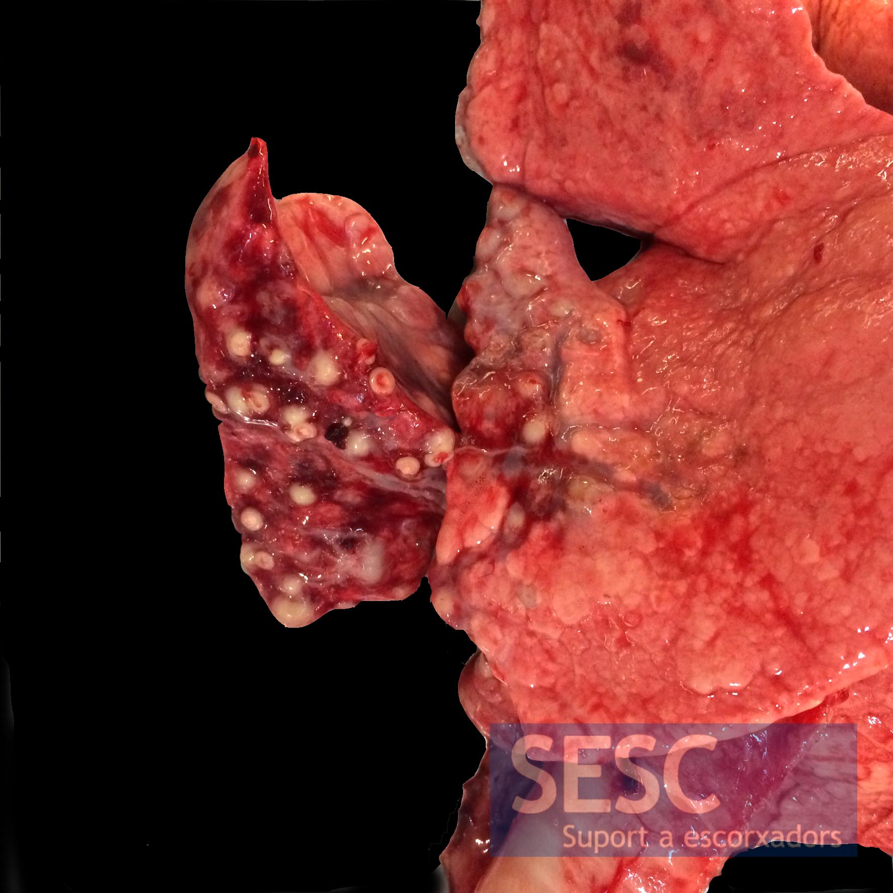
Apical lung lobe area with multiple mineralized spherical granulomatous lesions.
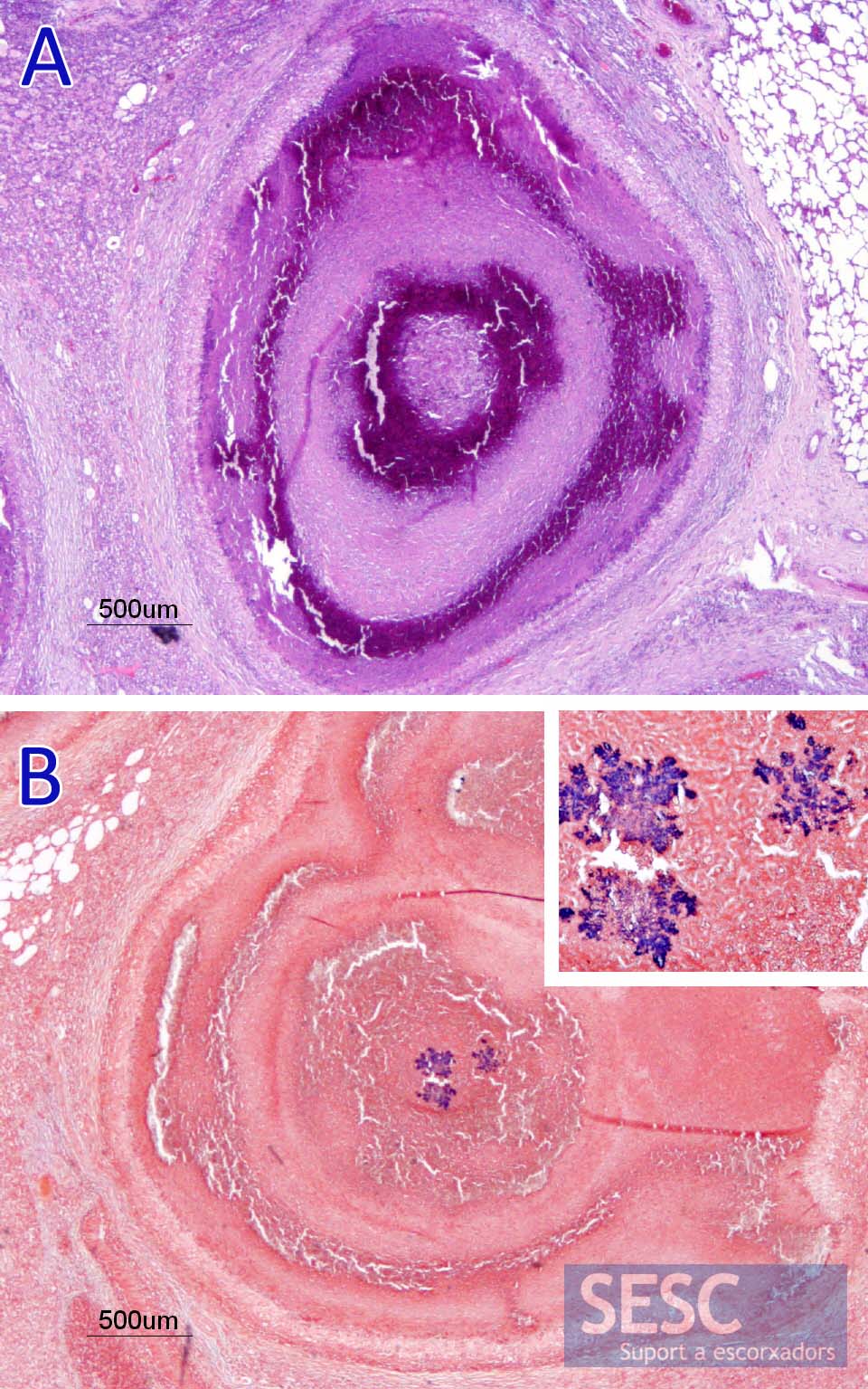
A: Lung granuloma, notice the concentric layers of necrotic mineralized material (strong lilac) surrounded by macrophages and fibrosis. B: Gram stain to visualize growth of gram positive bacteria (dark blue, at higher magnification in the inset) in the center of one of the granulomas. The growth morphology is characteristic of Corynebacterium pseudotuberculosis.
