Easter born Lambs II
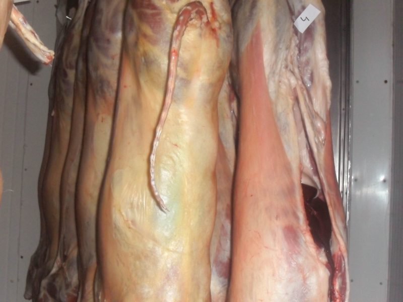
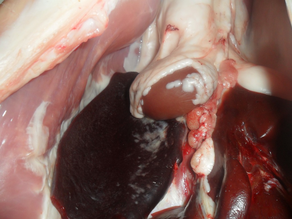
, moderate splenomegaly and hepatomegaly (see images 1 and 2). A similar case was discussed earlier in this forum: Link to entry Easter born lambs.
Histopathological study evidenced increased cellularity in the white pulp of the spleen with images of erythrophagocytosis. Most animals studied had mild inflammatory lesions in the hepatic parenchyma and erythrophagocytosis in Kupfer cells.
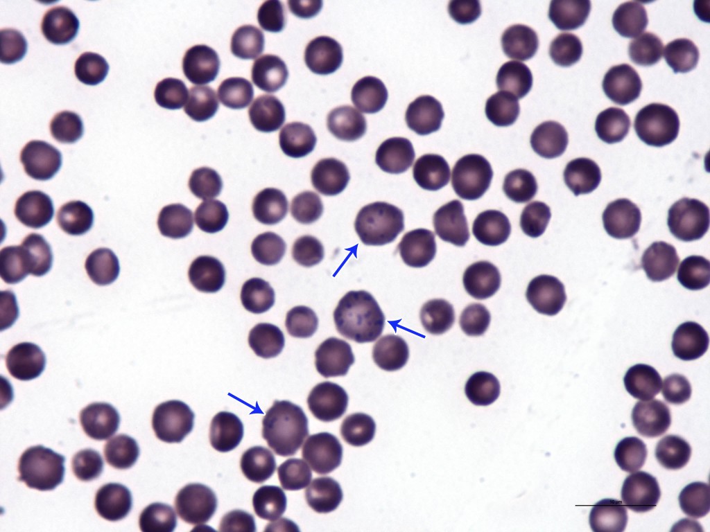
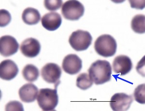
Blood smears were studied of most animals, images indicative of the presence of regenerative anemia and cocci shaped structures on the surface of erythrocytes were observed (see pictures 3 and 4).
Together, macro and microscopic findings are suggestive of an infection by Mycoplasma ovis (formerly known as Epheritrozoon ovis).
Finally a genus Mycoplasma PCR was performed in an attempt to confirm the presence of this bacterium using frozen liver samples. The result, however, was negative. It should be noted thtat the PCR was performed in only three animal samples and the ideal sample for this technique is blood. Thus, despite the negative PCR result, we do not exclude the diagnosis of Mycoplasma ovis.


