BLOG
SESC case archive


19/07/2011 | Kidney (porcine)
0

Widespread nodular lesions in a porcine carcass

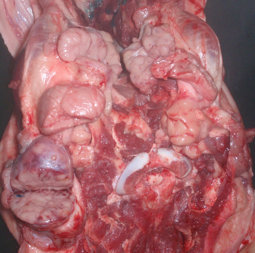
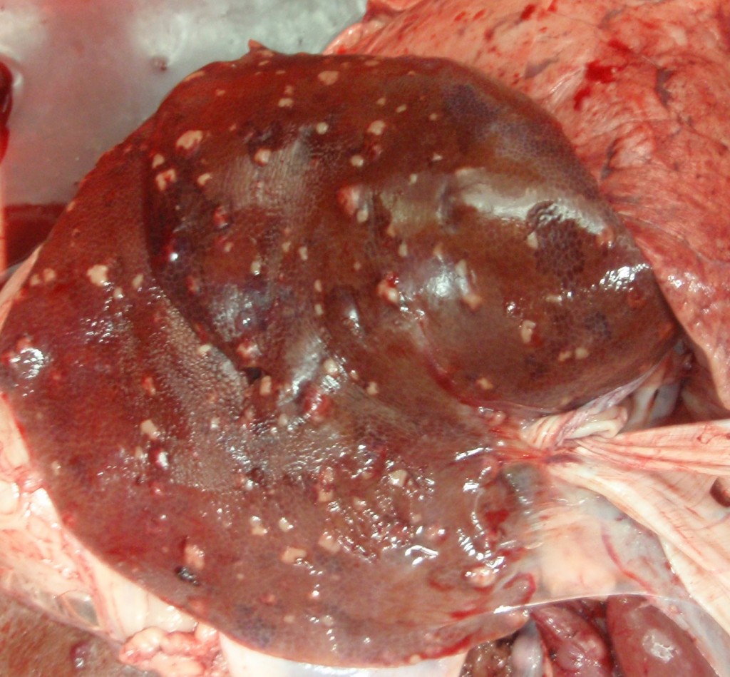
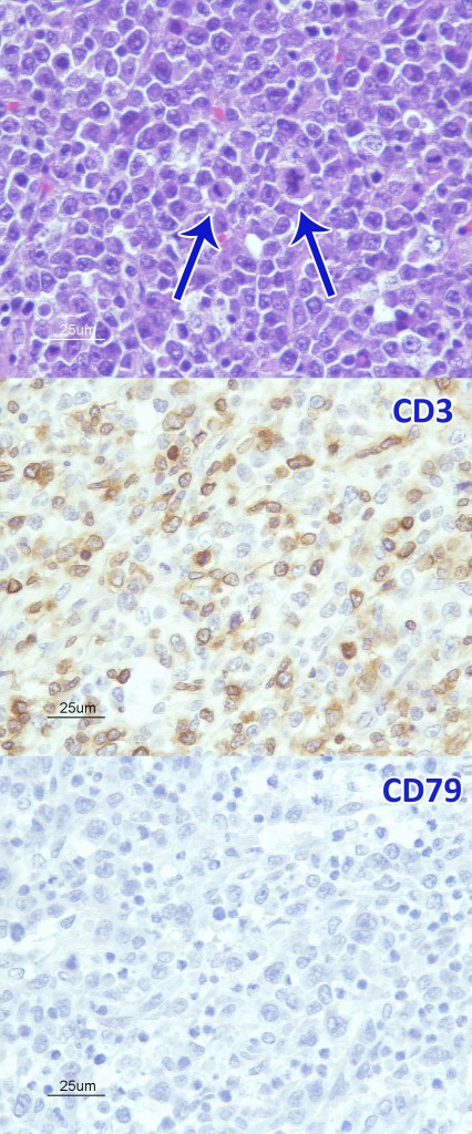
All the lesions observed (kidney, liver, heart and lymph nodes) were constituted by a single round cell neoplastic proliferation. The presence of lesions compatible with mycobacteriosis was ruled out. Immunohistochemical characterization allowed to classify the tumour as a T cell lymphoma.

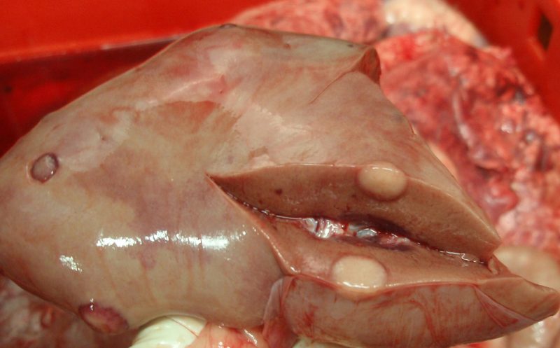
Nodular whitish multifocal renal lesions.

Lesions in the submandibular lymph nodes.

Nodular whitish multifocal hepatic lesions.

Histological image showing features of malignancy such as mitosis (arrows). Immunohistochemical markers: positive for T lymphocytes (CD3) and negative for B lymphocytes (CD79a), these results indicate that it is a T cell lymphoma.


Add Comment

Your email address will not be published. Required fields are marked *