

Traumatic erosive-ulcerative dermatitis in broilers (scabby hip)
A query was submitted from a poultry slaughterhouse: they frequently observe skin lesions on broiler carcasses, originating from farms of different origins, distributed over the lateral areas of the limbs and, occasionally, on the breast. The lesions have a linear morphology, with a brownish-yellow coloration, and the skin appears thinner and more translucent. This finding can lead to total condemnation of the carcasses as unfit for human consumption, although no lesions are observed in the viscera, nor any alterations in the underlying musculature.
Several affected carcasses were submitted for examination, and the slaughterhouse requested that we characterize the lesions in order to classify them properly.
At necropsy, the aforementioned lesions were observed in both scalded and unscalded carcasses, confirming the absence of visceral lesions and no involvement of the musculature.
Histopathologically, the lesions correspond to a loss of epithelium associated with crust formation composed of degenerated heterophilic leukocytes infiltrating the connective tissue of the dermis, with fibrin and some bacterial colonies.
In other areas, the lesions are milder and limited to focal accumulations of heterophils in the superficial layers of the epidermis.
Given the distribution and morphology of the lesions, the most probable explanation is that they correspond to scratches between animals (overcrowding during transport, piling due to high stocking densities, mounting behaviour, etc.).
The lesion can therefore be described as a traumatic erosive–ulcerative dermatitis. In English, this condition is referred to as scabby hip.
These are superficial, acute lesions, without alteration of the muscle quality, but with implications for animal welfare and potential economic losses due to organoleptic defects, since carcasses are commercialized with skin. It should be noted that the acute nature of the lesions indicates that the trauma occurred quite recently, during the final stages of fattening or even during animal transport. (EV)
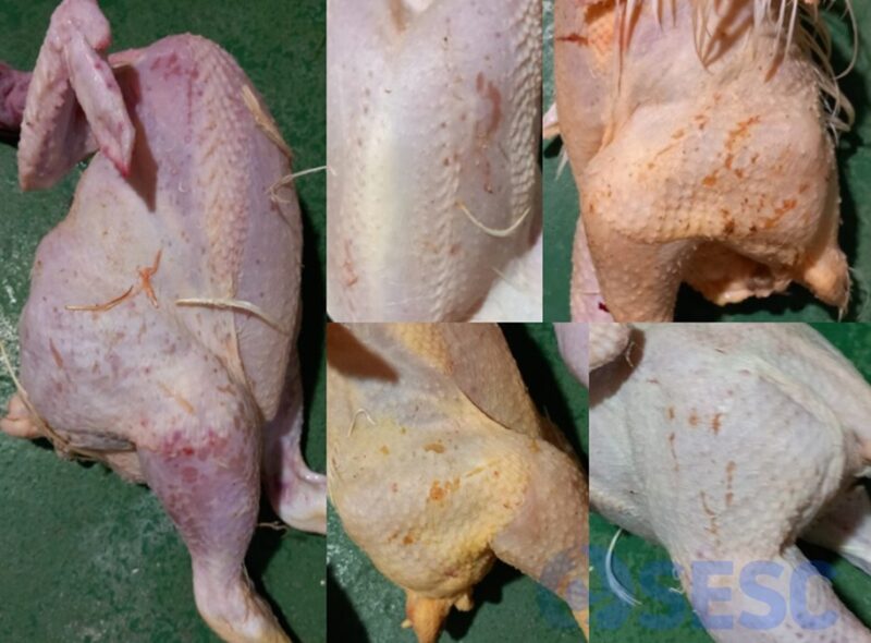
Broiler carcasses, skin. Several examples of skin lesions, often linear in morphology, yellowish-brown in color, observed mainly on the sides, thigh skin and breast.
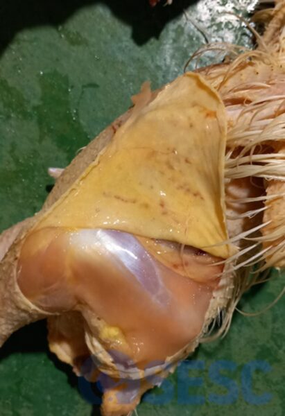
Broiler carcass, skin. When removing the skin, it is observed that the underlying musculature is not affected.
Below we show you images of three of the cases analyzed at SESC:
Case nº1
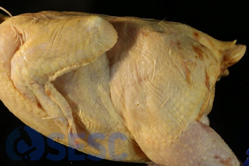
Broiler carcass nº 1, scalded. Erosive-ulcerative lesions in the dorsal region of the thigh.
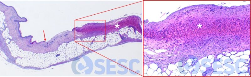
Histological image (H&E) of the lesions of carcass nº 1. A loss of epithelium can be observed (the red arrow marks the point where the epithelium is lost, which on the left appears normal). On the right, a crust (white asterisk) formed by an accumulation of degenerated heterophils can be observed, covering the connective tissue of the dermis.

Histological image (H&E) of the lesions of carcass no. 1. A loss of epithelium can be observed (the red arrow marks the point where the epithelium is lost) as well as accumulation of heterophils (asterisk) coinciding with the epidermal defect.
Case nº 2
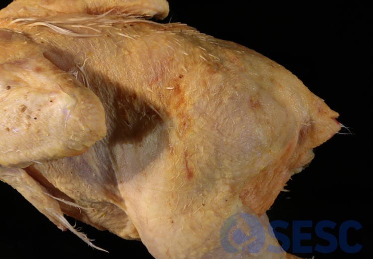
Broiler carcass nº 2, scalded. Erosive-ulcerative lesions in the dorsal region of the thigh.

Histological images (H&E) of the lesions of carcass nº 2. A loss of epithelium can be observed (red arrows mark the points where the epithelium is lost). The formation of crusts (white asterisks) formed by an accumulation of degenerated heterophilic leukocytes infiltrating the connective tissue of the dermis can be observed.
Case nº 3
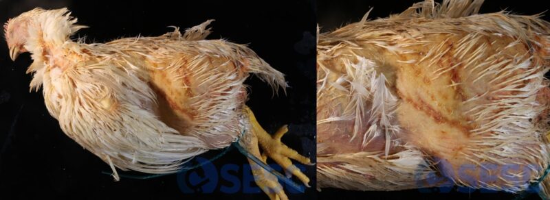
Broiler carcass nº 3, not scalded. Linear erosive-ulcerative lesions on the thigh (enlarged image in right panel).

Histological image (H&E) of the lesions of carcass nº 3. A loss of epithelium can be observed (the red arrow marks the point where the epithelium is lost). The formation of crusts (white asterisk) formed by an accumulation of degenerated heterophilic leukocytes infiltrating the connective tissue of the dermis can be observed.