

Bacterial endocarditis due to Moraxella lacunata in a bovine heart
We received an inquiry regarding a 10-month-old cross-breed male calf, in which, upon opening the heart, a multinodular lesion with an appearance reminiscent of a cauliflower was observed. When examining the sample, it was possible to observe an extensive lesion with a reddish coloration and a rough surface with a multinodular appearance, about 3cm in diameter, located in the mitral valve.
Histologically, it corresponded to a chronic thrombotic-septic proliferative lesion that caused a complete distortion of the architecture of the mitral valve. In the internal portion and at the subendothelial level, a very abundant amount of granulation tissue was observed; among which eosinophilic bright and radiated, amorphous material, surrounded by epithelioid macrophages was found along with viable and degenerated neutrophils and basophilic cellular debris (this finding is known as the Splendore Hoeppli phenomenon and is the result of the deposition of immunoglobulins around of a microorganism), in addition, areas of multifocal mineralization surrounded by macrophages and multinucleated foreign body giant cells were observed.
A Gram stain was performed, in which abundant Gram-negative bacterial colonies, which showed a tendency to form long coccoid chains, could be seen between the thick fibrin layers of the inflamed valve. A bacteriological culture was also carried out in which abundant growth of Moraxella lacunata was obtained.
Thus, a final diagnosis of proliferative thrombotic-septic valvular endocarditis caused by Gram-negative bacteria (Moraxella lacunata) was reached.
The genus Moraxella is composed of aerobic Gram-negative cocci. Fourteen different species of Moraxella have been isolated from various animals and humans, and several species exist as normal natural flora. Of these species, Moraxella lacunata can cause conjunctivitis, keratitis, endocarditis and otolaringitis in humans, but is rarely found in animal cases. In case of finding lesions like these, it is important to carry out a detailed inspection of the rest of the carcass and viscera, since it is common that fragments of the lesion, which will contain bacteria, can embolize and move via the bloodstream to other locations. (MC)
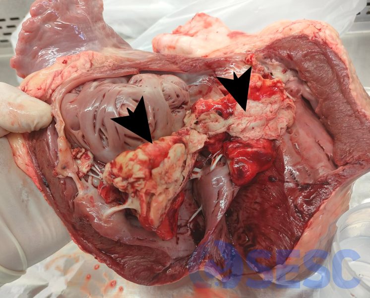
Bovine heart, mitral valve. Proliferative, chronic, whitish lesions in the shape of "cauliflower" approximately 3 cm in size.
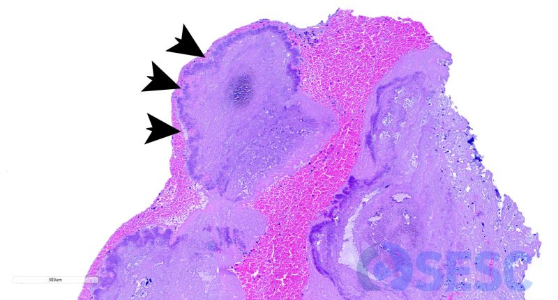
Histological image at low magnification, showing necrosis, inflammation, and bacterial colonies (black arrows) on the mitral valve.

Gram stain: clusters of coccobacillary bacteria with a fuchsia pink coloration (Gram-negative) are observed.
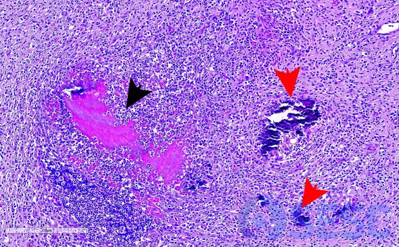
Hematoxylin-eosin: Splendore-Hoepli phenomenon (black arrow), multifocal areas of mineralization (red arrows), all surrounded by granulation tissue.