

Myopathies in chicken breasts
Myopathies are muscle defects related to hypoxia and oxidative stress, which ultimately involve the apoptosis of muscle cells and their replacement by fat and connective tissue. Their incidence is usually high in fast-growing broiler chickens, dedicated to being sold as cuts, where breast yield is especially important.
- White striping myopathy, is characterized by the presence of white stripes in the muscle fibers. This myopathy was the first to be detected and is quite common in the food industry.
- Wooden breast myopathy is the most studied for its significant impact on product quality. It is characterised by a hardening of the muscle, often accompanied by areas of pale color and petechiae.
- Spaghetti meat myopathy is the most recently identified. It is characterized by the loss of cohesion of the muscle fibers, which separate, giving the meat a spaghetti-like appearance.
All of these myopathies present similar lesions in a histological section, including the infiltration of white blood cells and the death of muscle cells, accompanied by infiltration of connective tissue, adipose tissue and water. In the case of white striping, it is mainly adipose cells that occupy the space of the muscle fibers. On the other hand, wooden breast and spaghetti meat show thicker connective tissue. In the cases of wooden breast, the collagen fibers are more compact, while in spaghetti meat they are smaller and immature, thus explaining the lack of cohesion between the muscle fibers.
Entry written by our colleagues from IRTA - Food Industries (Monells).
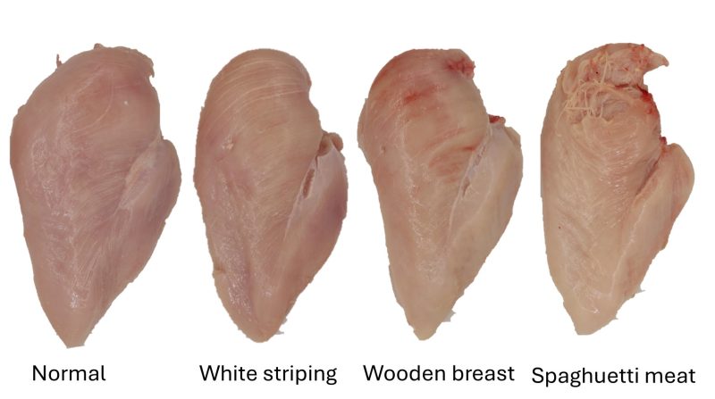
Aspect of the myopathies observed in chicken breast.

Histological appearance of a normal muscle (left), one with wooden breast (center) and one with spaghetti meat (right) in a collagen stain (red). In the lower panel observed with polarized light. m: muscle fiber, m-n: degenerated muscle fibre, p: perimisium. Image: Sanden et al 2021.