Chronic peritonitis in pig carcasses
In Case 1 both the thoracic and the abdominal cavity were involved with a rather reddish exudate (haemorrhage). Case 2 was restricted to the abdominal cavity and it was characterised by adhesions between viscera and the deposited material had a whitish color. The animal had a noticeable loss of weight.
Both macroscopic appearance and subsequent histopathological study are consistent with chronic diffuse peritonitis with an important fibrous component, probably due to chronic fibrinous peritonitis. At this stage of the disease a microbiological analysis would be usless as the causative agent of peritonitis is probably no longer present.
The most common causes of fibrinous poliserositis in swine are: Haemophilus parasuis, Streptococcus suis, Escherichia coli and Mycoplasma hyorrhinis.
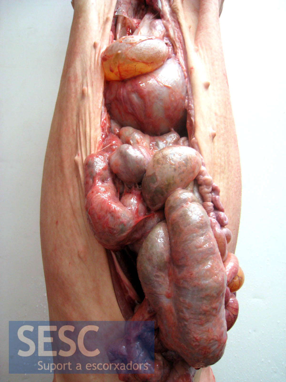
Case 1, deposition of fibrous material on the abdominal serosal surfaces.
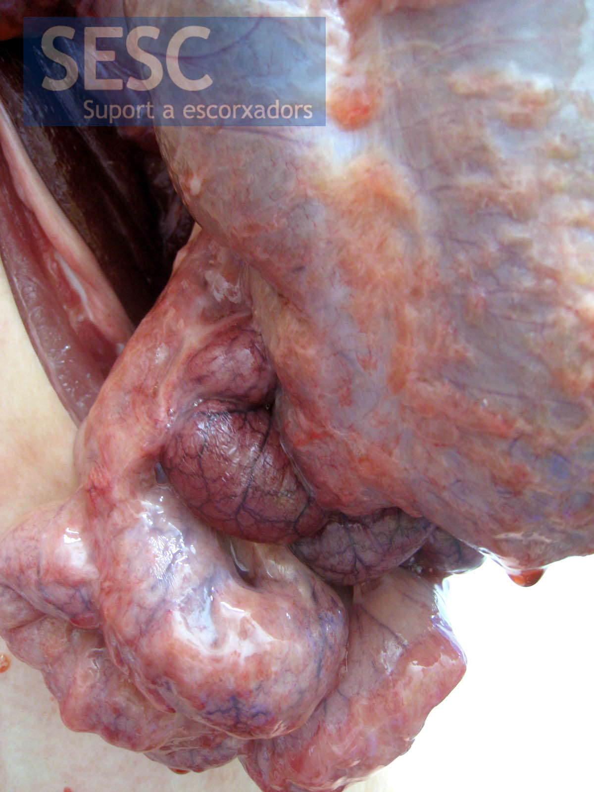
Case 1, detail of the inflammatory exudate.
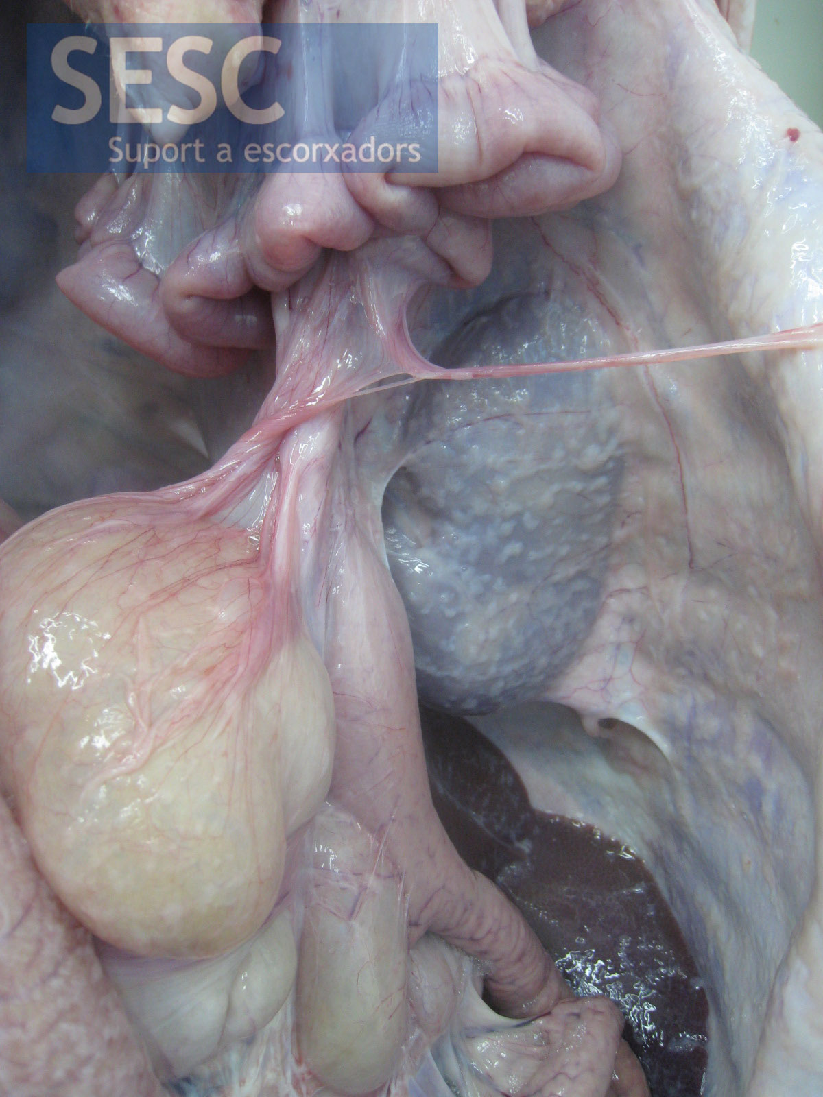
Case 2, whitish deposits of fibrous material were observed.
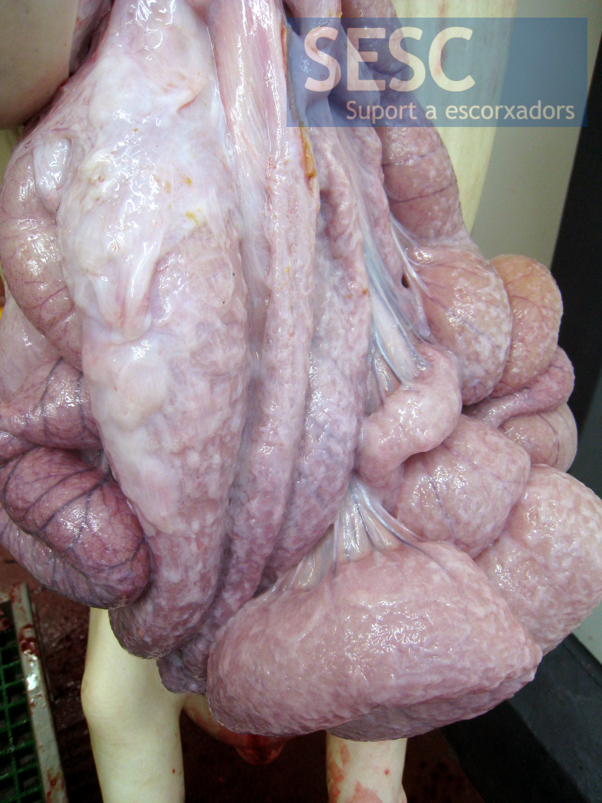
Case 2, the viscera had adhesions between them and were covered by abundant fibrous tissue.

4 comment(s)
Should carcasses like that be condemned since they hold no health risque after removal of the affected intestines?
It will depend on a fine assessment of the meat inspector of each particular case of course, whether it s a local (only peritenoeum) or more extended lesion (polyserositis), the type of inflammation, involvement of lymph nodes and other viscera… but indeed a resolved fibrous peritonitis dose not pose any risk to public health. Provided that the peritoneum abdominal viscera and intestines are removed, the meat should be safe.
Buena tarde, hemos tenido varios casos de este tipo en rastro, ¿que es lo que deberia hacerse con estas canales, se pueden mandar a retrabajo o es decomiso automatico total de canal y viscera?
El inspector veterinario debe hacer una evaluación precisa de cada caso. En función de la extensión y gravedad de la lesión, si afecta solo el peritoneo o también otras serosas (poliserositis), si estan afectados los nódulos linfátics u otras visceras. Lo cierto es que una canal con peritonitis fibrosa crónica, siempre qeu sean debidamente retirados el peritoneo, las visceras abdominales y los intestinos la carne no debiera suponer ningún riesgo para la salud humana.