Detection of swine fever lesions in slaughterhouses and game meat processing facilities
African swine fever (ASF) and classical swine fever (CSF) are two notifible viral diseases of domestic pig and wild boar that can cause high mortality in pig farms, and clinically presenting mainly fever and hemorrhages.
Although these viruses are not present in Catalonia, an eventual entry of the disease cannot be ruled out. A possible route would be through the entry of infected wildlife (wild boar) as in eastern European countries ASF and CSF are endemic. Source: WAHIS, OIE.
Slaughterhouse surveillance can be a key tool in detecting the presence of lesions of these diseases. Therefore, especially in game meat processing facilities and in swine slaughterhouses, it is necessary to remain alert to the presence of swine fever compatible lesions.
For example, in cases like this one we published a while ago: Hemorrhagic diathesis in a pig carcass. Lesions were observed that could be compatible with either classical or African swine fever.
In the event of a suspected case, refrigerated lymph node and tonsil samples should be submitted to SESC to perform a PCR and discard the presence of viruses.
A while ago, we reviewed the lesions caused by African swine fever virus:
It is important to keep in mind that, in wild boar carcasses, it is very easy to observe blood resorption on the lymph nodes as a consequence of hemorrhages caused by firing.
In this post we show a few images of lesions caused by the Classical Swine Fever virus obtained from experimental infections at the IRTA-CReSA OIE CSF Reference Laboratory.
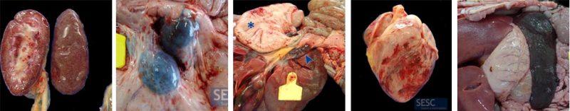
Petechiae (hemorrhages smaller than 1mm in diameter) on the kidneys.
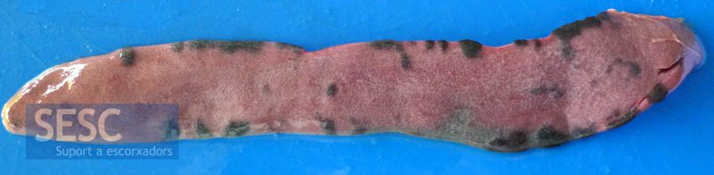
Peripheral areas of infarction on the spleen.
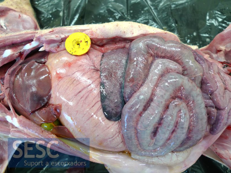
Splenomegaly (with peripheral infarction).
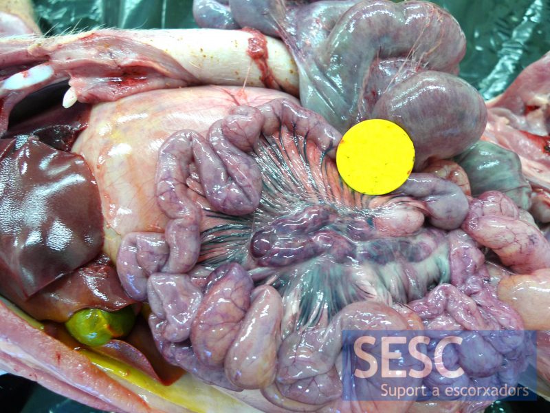
Hemorrhagic (mesenteric) lymph nodes.
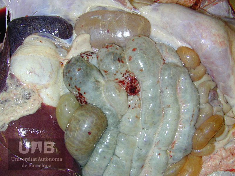
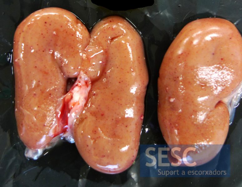
Hemorrhagic lesion in the intestinal wall and renal petechiae, in a field CSF case.
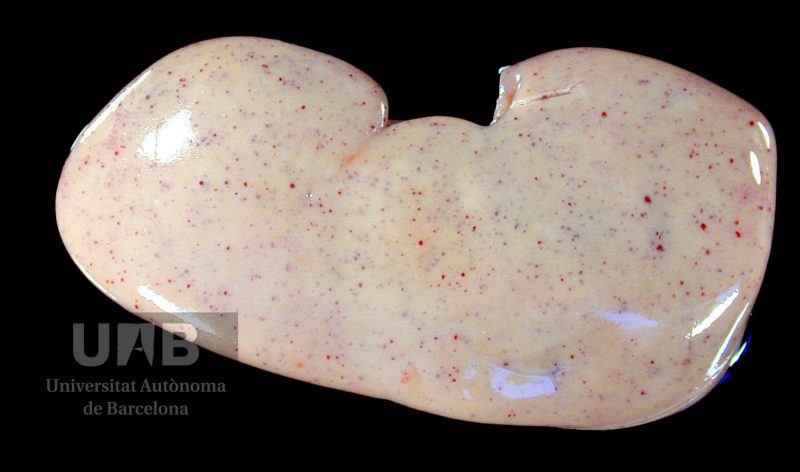
Detail of the renal petechiae of the field CSF case shown above.
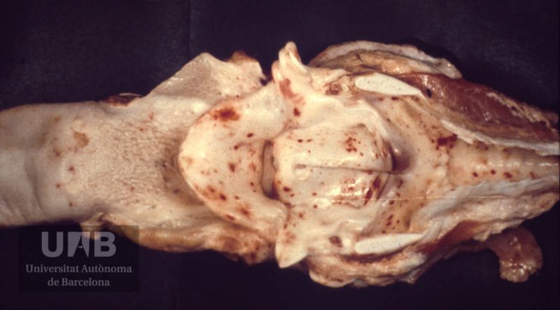
Petechiae in the trachea and glotis in a CSF field case.
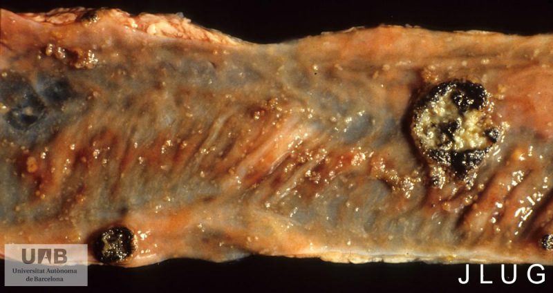
Button shaped ulcer in the colon of a CSF field case. Source SDPV image archive.
