Gangrenous dermatitis in a turkey by S. aureus
We received an inquiry regarding a 127-day-old hybrid turkey, which showed extensive cutaneous lesions, with necrotic appearance, showing reddish discolouration between ulcerated areas and a gelatinous appearance of the subcutaneous tissue. Additionally, the animal presented a low corporal condition, as well as bursitis on the right tarsal joint.
We received fresh skin samples and proceeded to their histopathological study. The skin lesions observed corresponded to focally extensive areas of ulceration and substitution of the epidermis with abundant necrotic debris, degenerated leucocytes, erythrocytes, and fibrin (haemorrhagic and necrotizing dermatitis, multifocal and extensive, subacute and severe). Additionally, numerous cocci could be observed adhered to the necrotic debris, which under a gram stain appeared to be gram-positive.
Given the histological appearance, the main suspicion consisted of a gangrenous dermatitis (caused by clostridium or staphylococci, the latter being the main suspicion due to the observation of gram-positive cocci). These lesions usually have a traumatic origin, that leads to bacterial contamination of the skin and development of the necrotic lesions. To confirm such suspicion, we submitted samples to the microbiological service, and the culture yielded (aside from minor contamination) abundant colonies of coagulase positive Staphylococcus. (AC)
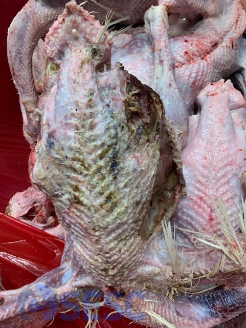
Multifocal cutaneous lesions at the dorsal region of the animal. Extensive areas of ulceration can be seen, accompanied of a gelatinous appearance of the subcutaneous tissue (edema).
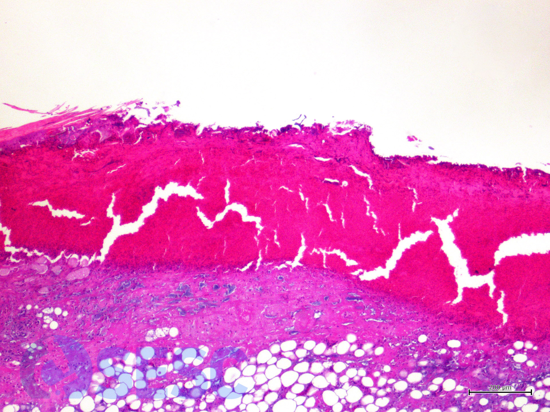
Histologic detail of the lesion. A loss of the epidermis can be seen, replaced by an accumulation of cellular debris, degenerated leukocytes, erythrocytes and fibrin (necrotizing and haemorrhagic dermatitis).
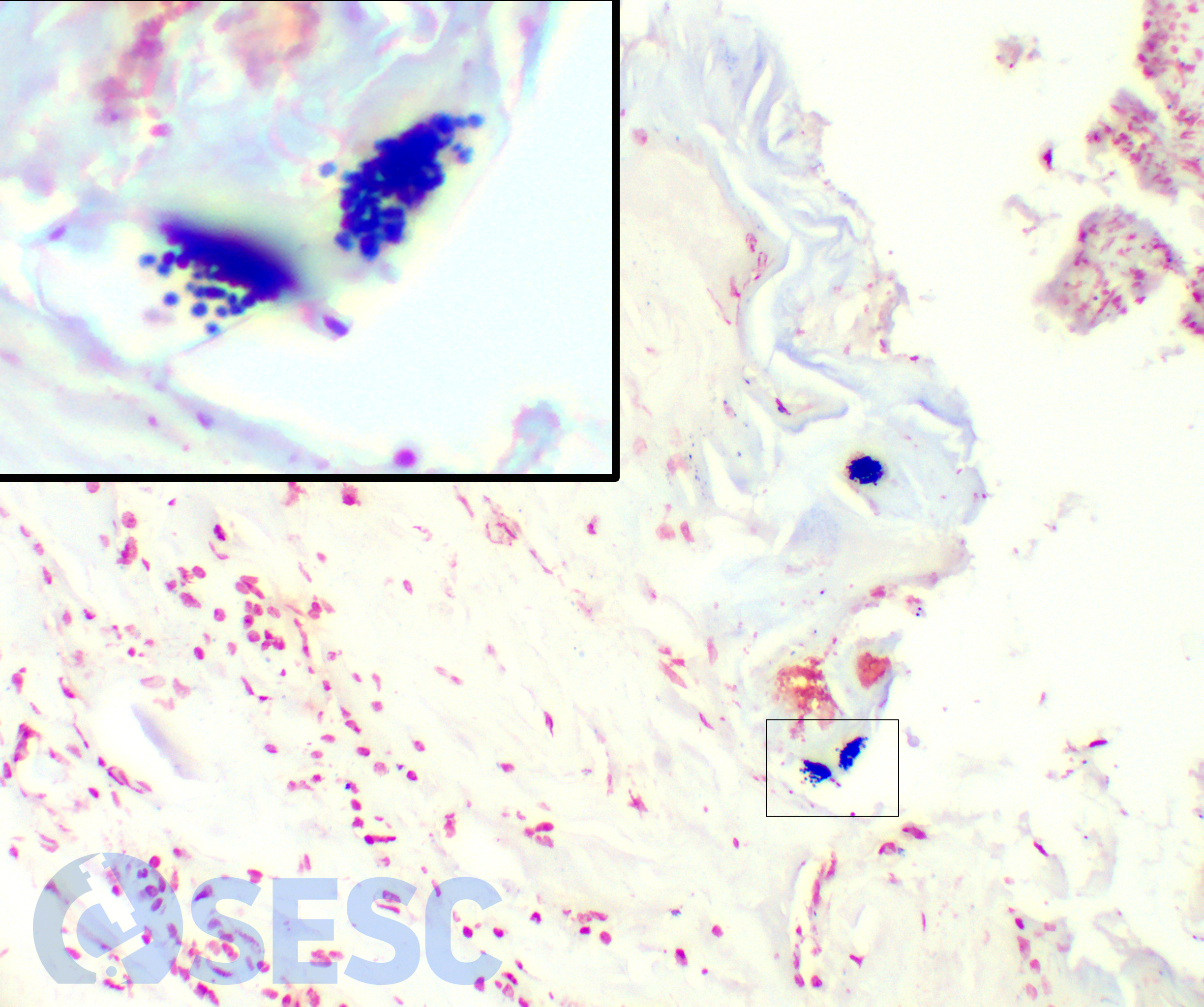
Histologic detail of the lesion under a Gram stain. Gram-positive cocci can be seen associated to the lesion crusts.
