Granulomatous lymphadenitis in a wild boar
A meat inspector form a an establishment processing game meat subitted samples of tracheobronchial and prescapular lymph nodes froma a wild boar carcass (Sus scrofa). The lymph nodes were slightly increased in size and hardened. When sectioned, mineralized and whitish lesions were obsereved.
Histopathology showed large areas of necrosis, mineralization and the presence of a granulomatous inflammatory infiltrate (predominantly macrophages) with presence of multinucleated giant cells. These lesions are compatible with tuberculosis.
The Ziehl Neelsen stain did not allow the identification of acid-alcohol resistant bacilli, but microbiological culture allowed to obtain a growth which, by means of a PCR, was identified as Mycobacterium caprae. This is the main cause of TB in goats, and can also infect other species such as cattle, sheep and also human beings.
In the wild boar the location where more frequently TB lesions are observed are the submandibular lymph nodes.The appearance of the lesions varies, from small mineralized sand grain-like points to a marked increase in size with whitish, more or less hardened, granulomatous lesions.
Game meat processing establishments are an important point for monitoring outbreaks of tuberculosis in wildlife.
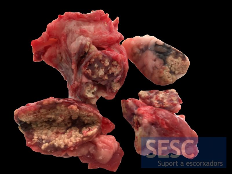
Granulomatous lymphadenitis in the tracheobronchial lymph nodes of a wild boar.
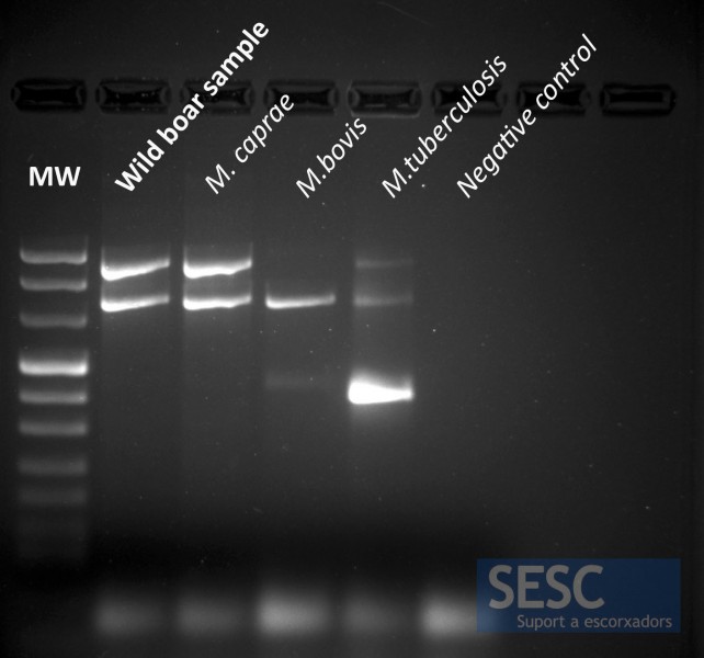
PCR gel image where one can see that th eband for the culture of the studied wild boar sample matches the M.caprae band pattern.
