Hepatic abscesses in an adult bovine
In a 37-month-old cattle carcass belonging to the brown Pyrenees breed, multiple nodules of 2 to 3 cm in diameter were observed in the liver, with a soft, yellowish content with purulent appearance. When removing the liver with the rest of the viscera, adhesions to the rumen were observed and an abscess with blackish fluid content ruptured.
The histopathological study revealed that the abscesses observed showed a lytic necrosis centre with abundant coccoid bacteria, surrounded by a rim of neutrophils that was in turn delimited by foamy macrophages, fibrosis and lymphoplasmacytic cells, thus confirming that the lesion consists of subacute multifocal and generalized hepatic abscesses.
The sample was also sent to the microbiology service, where Trueperella pyogenes was cultivated in large quantities, as well as colonies of Staphylococcus sp.
Hepatic abscesses can have multiple origins, but not all are equally likely, the casuistry being highly variable:
- It is assumed that the main cause of hepatic abscesses is hematogenous spread through the portal vein, by bacterial translocation from the draining viscera (digestive system). In this sense, the usual bibliography associates calf ruminitis (due to ruminal acidosis) as the most frequent cause in the secondary development of liver abscesses caused by Fusobacterium necrophorum.
- Another important but less frequent possibility of hepatic abscesses is an ascending infection through the umbilical vein in animals that have suffered from neonatal omphalitis. In these cases, the gross lesions could be very similar to the previous modality. However, in this specific case of a 3-year-old animal, this possibility is considered negligible.
- Other causes of purulent lesions in the liver are also described, although their prevalence is much lower than the previous ones. These possibilities are as follows:
- Septicaemia: bacteria can access the liver through the hepatic artery. In this case, this option was ruled out due to the absence of lesions compatible with a systemic condition.
- Biliary: ascending infection of biliary origin is described in the literature as a cause of purulent hepatitis. In our experience, in these cases the gross lesions don’t feature a generalized multifocal dissemination like the one observed. Additionally, the characteristics of these lesions respond more to a purulent cholangiohepatitis than to hepatic abscesses.
- Parasitosis: much less frequent is the finding of liver purulent lesions of parasitic origin. In any case, involution of cestode cysts that evolve (becoming contaminated) towards secondary suppurative lesions are described.
- Direct extension: practically irrelevant from the point of view of the casuistry, the extension of an inflammatory process adjacent to the liver could also be considered as a cause of purulent lesions (such as traumatic reticulitis).
In conclusion, in this case the age of the animal, the generalized multifocal nature of the abscesses, and the absence of other systemic lesions suggest a portal hematogenous spread; however, it is a common presentation in calves, but very rare in adult bovines. Another uncertain possibility in this case is that the hepatitis detected was related to the abscess described by the inspectors (broken when removing the viscera).
Another interesting aspect that is probably worth mentioning is the bacteria involved in this injury. The literature initially attributes the origin of liver abscesses to the arrival of Fusobacterium necrophorum via the portal vein, but the commonly observed lesions and microbiological results almost never coincide with this statement. This contradiction is explained by speculating that the original necrotizing necrobacillosis lesion is secondarily contaminated with pyogenic bacteria, the most frequently isolated being Trueperella pyogenes. (AC)
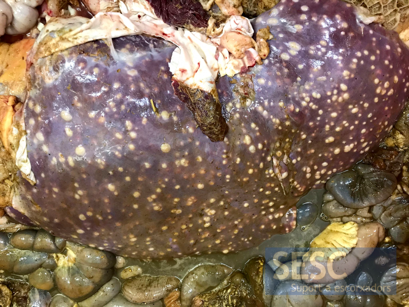
Liver and other viscera, in which the multiple hepatic abscesses can be seen throughout the serosa surface.
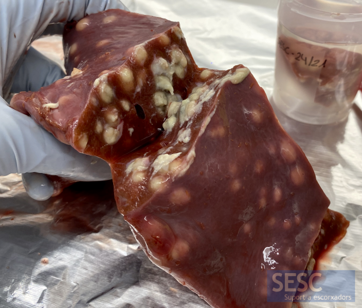
Cut section of the liver, where the hepatic abscesses can be seen through the entire parenchyma.

1 comment(s)
Buen articulo,