

Systemic necrobacillosis in a calf
An inquiry was received regarding a carcass of an 11-month-old Holstein calf. No relevant abnormalities were observed during the ante-mortem inspection; however, post-mortem examination revealed multiple whitish nodular lesions with a necrotic appearance, distributed in a random multifocal pattern across several organs (liver, spleen, kidney, diaphragm, heart, and skeletal muscle). These lesions showed marked size variability (ranging from 0.5 to 3 cm in diameter) and extended deeply into the parenchyma. Additionally, the carcass showed generalized findings consistent with septicemia, such as renal petechiae. Based on all observed findings, the carcass was declared unfit for human consumption.
The submitted samples from liver, skeletal muscle, and heart showed multifocal to coalescing, whitish-yellowish nodular lesions. The kidney showed multiple irregular whitish-yellow areas in the cortex, as well as small punctate reddish foci.
Histopathological examination revealed severe, subacute, multifocal to coalescing necrotizing hepatitis; severe, subacute, transmural necrotizing myocarditis and endocarditis; severe, subacute, focally extensive necrotizing nephritis; and severe, subacute, multifocal to coalescing necrotizing myositis, all of them with the presence of intralesional bacterial colonies.
Gram staining revealed that the observed bacteria had a filamentous morphology and were Gram-negative. Microbiological culture of the lesions yielded abundant colonies of Fusobacterium necrophorum in all evaluated organs.
The observed lesions, together with the bacterial isolation, were suggestive of systemic necrobacillosis.
The primary focus of infection could not be determined. In cattle, this microorganism is part of the normal microbiota of the digestive tract and is usually associated with localized processes, mainly hepatic abscesses secondary to a) ruminitis—frequently related to ruminal acidosis in animals fed high-grain diets—or b) infectious pododermatitis, one of the main causes of lameness in beef and dairy cattle. However, no disseminated systemic presentations have been described in this species.
In human medicine, F. necrophorum is the classical etiological agent of Lemierre’s syndrome, an entity characterized by septic thrombophlebitis of the internal jugular vein secondary to an oropharyngeal infection, with hematogenous dissemination and formation of septic emboli, mainly in the lungs. Similarly, in the bovine case described, the multifocal distribution of necrotizing lesions in several organs also suggested hematogenous dissemination with the formation of septic emboli (ML).

Sectioned liver with multiple intraparenchymal nodules of necrotic appearance.

Another liver section, in which the necrotic areas showed irregular morphology.

View of the endocardium with multiple whitish necrotic areas.
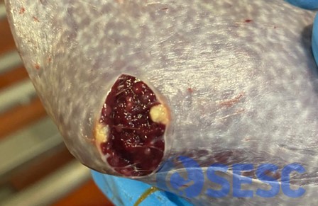
Sectioned spleen with a whitish intraparenchymal nodule.

Kidney section showing a whitish-yellow area in the renal cortex. Generalized, small millimetric reddish spots (petechiae) were also observed.

Examples of the lesions observed in skeletal muscle.

Histological section of skeletal muscle showing a focally extensive area of lytic necrosis affecting muscle fibers.

Histological section of the kidney showing a focally extensive area of lytic necrosis replacing glomeruli and tubules in the renal cortex.