Hydatidosis in a sow carcass
In a 2-year-old, hybrid breed, sow carcass, multiple vesicular, whitish, variable-sized lesions, between 2 and 4 cm in diameter, were seen in the liver parenchyma.
Samples of the cysts were sent to the parasitology laboratory where the presence of protoescolices was observed, thus classifying the lesion as a fertile unilocular hydatid cyst (or Cystic echinococcosis). The protoescolices form what is known as hydatidic sand which is found inside the vesicles. Protoscolices are not visible to the naked eye, a microscope is required to identify them. It is a platyhelmint parasite, class of cestodes, Taenidae family and the genus Echinococcus. This information allows us to identify it as Echinococcus granulosus sensu lato.
The definitive host of E. gransulosus is the dog, who has the taenia in its small intestine and excretes embryonated eggs in its scat. The intermediate hosts can be various, usually sheep, but we can also find it in goats, cattle and pigs, among others, as well as people, as it is a zoonosis. When these intermediate guests ingest the eggs, the oncospheres eclose, they penetrate through the walls of the intestine and travel to the liver or to the lungs where hydatidal cysts develop. The dogs are infested by swallowing the offal with these cysts. To read more about the E. granulosus cycle click here.
The taxonomy of these parasites is currently based on molecular biology techniques that differentiate different genotypes within E. granulosus senso lato , among which we emphasize those that are zoonotic (T.Romig et al. 2015 Veterinary Parasitology):
- E. granulosus senso stricto (genotypes G1-3), usually transmitted by sheep.
- E. ortleppi (G5) transmitted by cattle.
- E. canadiensis that includes:
- E. intermedius (G7) transmitted by pigs and (G6) transmitted by camels.
- E. borealis (G10) transmitted by the cervix.
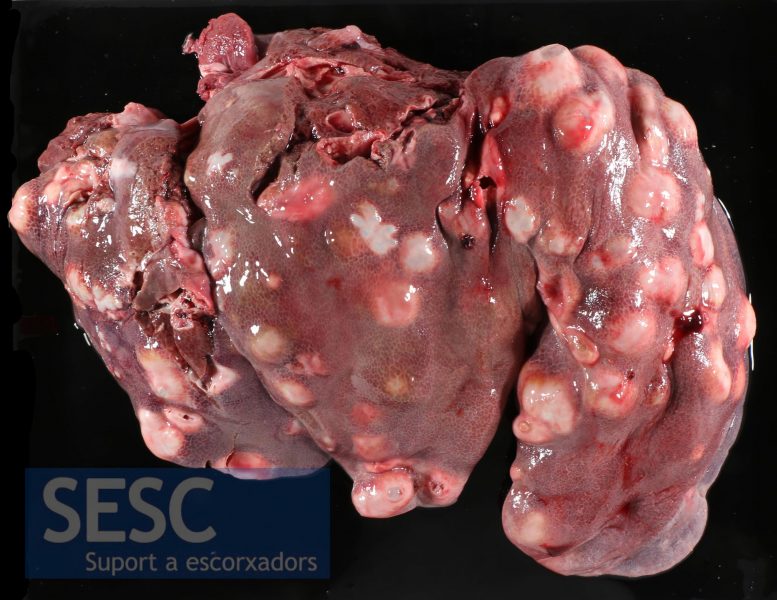
Sow liver with multiple hydatid cysts.
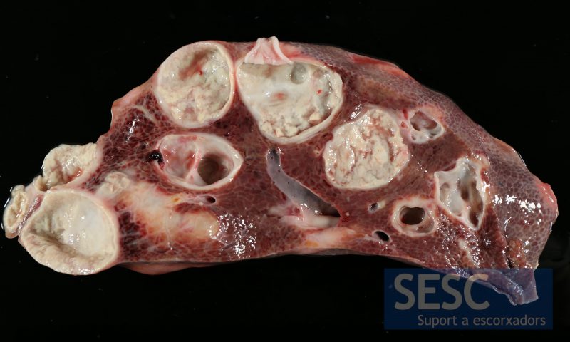
Liver section showing the inside of the vesicles.
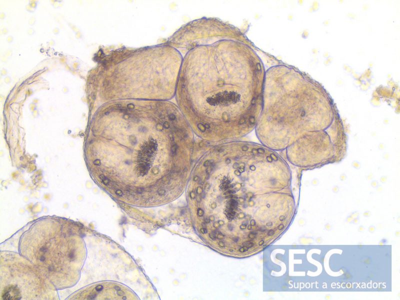
Within the vesicles protoescolices of E.granulosus (s.l.) were identified.
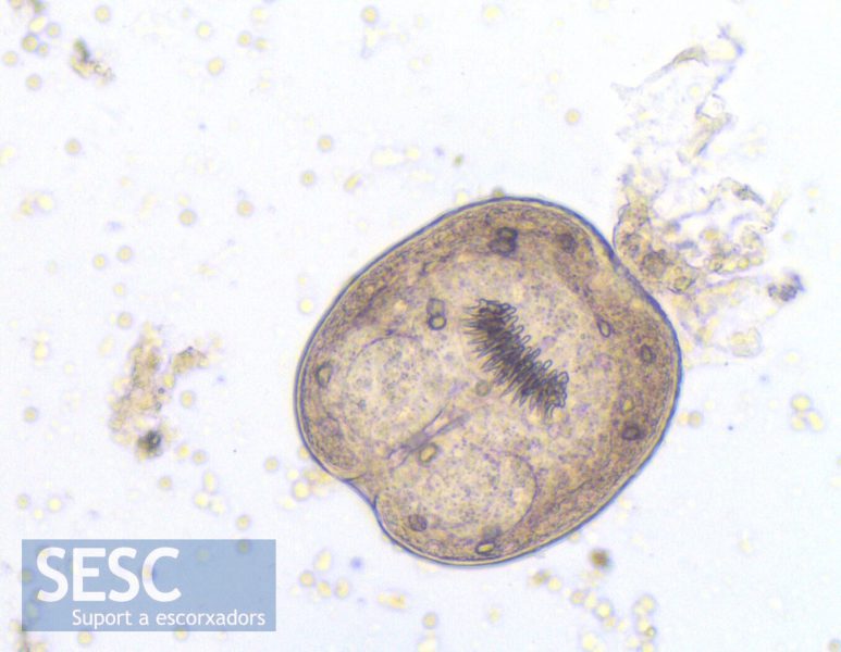
E.granulosus (s.l.) protoescolex.
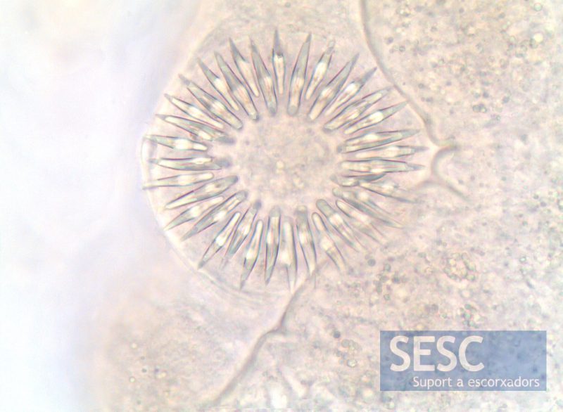
Detail of the hook crown in E.granulosus (s.l.) rostellum.
