Infection with African horse sickness virus (AHSV)
African horse sickness (AHS) is a disease caused by an RNA virus of the genus Orbivirus (family Reoviridae), closely related to bluetongue virus and epizootic haemorrhagic disease virus.Nine different serotypes have been described. The disease primarily affects horses; donkeys and mules are less susceptible, and zebras act as the main sylvatic reservoir. Dogs may develop disease after ingesting infected offal. AHS is not a zoonosis. Transmission occurs via biting midges (Culicoides spp., Diptera). AHS is listed by the World Organisation for Animal Health (WOAH) as a notifiable disease, and in the European Union it is included among Category A diseases under the Animal Health Law (Commission Delegated Regulation (EU) 2020/687).
The disease has been eradicated from the EU. Spain achieved AHS-free status in 1993, after an outbreak in 1985 originating from the importation of zebras in Madrid spread across the Iberian Peninsula, Portugal, and Morocco. In contrast, in sub-Saharan African countries the disease remains endemic.
Detection at slaughterhouses is less likely than clinical detection, given the acute course of the disease. The virus initially replicates in macrophages and dendritic cells, followed by viraemia and secondary replication in endothelial cells, leading to severe vascular damage. Consequently, marked increases in vascular permeability (oedema) and haemorrhages occur, particularly in the lungs, heart, pleura, and pericardium. The immune response also contributes to vascular damage. The following table describes the clinical forms and associated lesions:
Pulmonary form (“dunkop”)
-
Case fatality: ~95% (peracute in non-immune horses)
-
Clinical signs: fever, dyspnoea, tachypnoea, spasmodic coughing, abundant serofibrinous or frothy nasal discharge (often post-mortem).
-
Lesions: pulmonary oedema (failure of lung collapse at necropsy, usually absent in slaughtered animals); hydrothorax (yellowish exudate coagulating on contact with air); lymphadenomegaly of tracheobronchial and mediastinal lymph nodes with petechiae on the capsule; haemorrhages in epicardium and endocardium.
Cardiac form (“dikkop”)
-
Case fatality: ~50%
-
Clinical signs: fever, dyspnoea, cyanosis.
-
Lesions: subcutaneous oedema (yellowish, gelatinous) of the head and neck (supraorbital fossa, eyelids, tongue, intermandibular space), thorax and shoulders; conjunctivitis; petechiae on conjunctival and oral mucosa; hydropericardium; pulmonary congestion.
Mixed form
-
Case fatality: ~70%
-
Most common form.
-
Combination of pulmonary and cardiac signs and lesions.
Horse sickness fever
-
Usually subclinical.
-
Occurs in immune animals infected with heterologous serotypes or in resistant equids (zebras).
Differential diagnosis: Lesions are non-specific, although oedema in the supraorbital fossa is highly characteristic. Equine infectious anaemia (Retroviridae), arboviral encephalitides, Hendra virus infection (Henipavirus), equine viral arteritis (Arterivirus), equine influenza (Orthomyxoviridae), piroplasmosis/babesiosis and purpura haemorrhagica.
Diagnosis: If compatible clinical signs are detected at ante-mortem inspection, or suggestive lesions are found (notably in deaths during transport), confirmation is obtained by RT-PCR from anticoagulated whole blood (EDTA is recommended; heparin inhibits PCR) or from spleen, lung, or thoracic lymph nodes. The Spanish Reference Laboratory for African horse sickness is the Central Veterinary Laboratory (LCV) in Algete, Spain. (EV)

Horse. Palpebral oedema and conjunctival hyperaemia. Source: MAPA (Spanish Ministry of Agriculture, Fisheries and Food).

Horse. Periorbital and conjunctival oedema. Source: Dr Camilla Weyer.
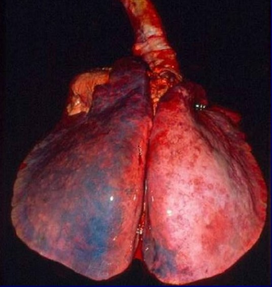
Horse. Lung. Failure of pulmonary collapse due to oedema. Source: USDA.
Horse. Lung. Marked interstitial (interlobular) pulmonary oedema. Source: PIADC.
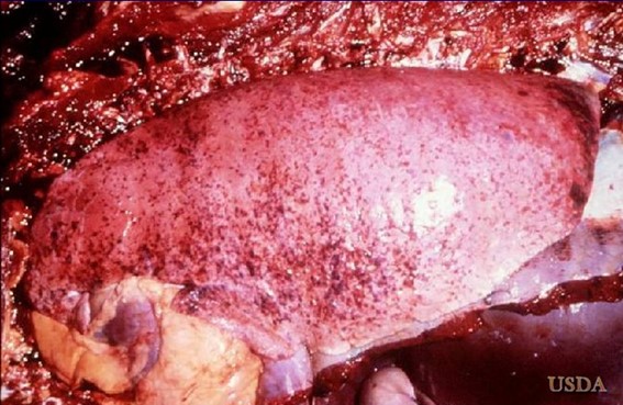
Horse. Lung. Petechial haemorrhages on the visceral pleura. Source: USDA.
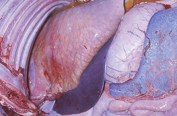
Horse. Heart. Hydropericardium. Source: PIADC.

Horse. Heart. Petechial haemorrhages on the endocardium. Source: PIADC.
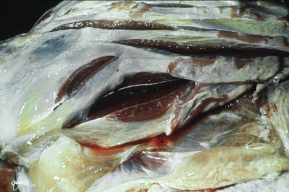
Horse. Skeletal muscle. Marked intermuscular oedema. Source: PIADC.
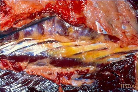
Horse. Skeletal muscle. Marked intermuscular oedema. Source: USDA.
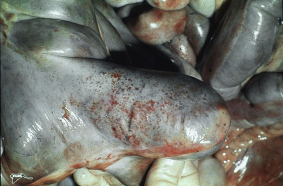
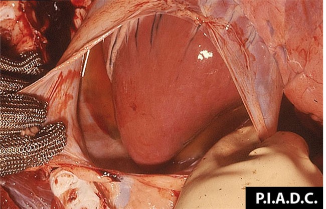
Horse. Cecum. Petechiae on the serosa of the cecum. Source: PIADC.
