Injection site myositis and panniculitis
We received an inquiry regarding 21, 12-months old, Montbeliard breed calves showing a focalized inflammatory process located in the right cervical area, affecting the braquiocefalic muscle. The localization and appearance of the lesions were compatible with lesions derived from a parenteral pharmacological administration.
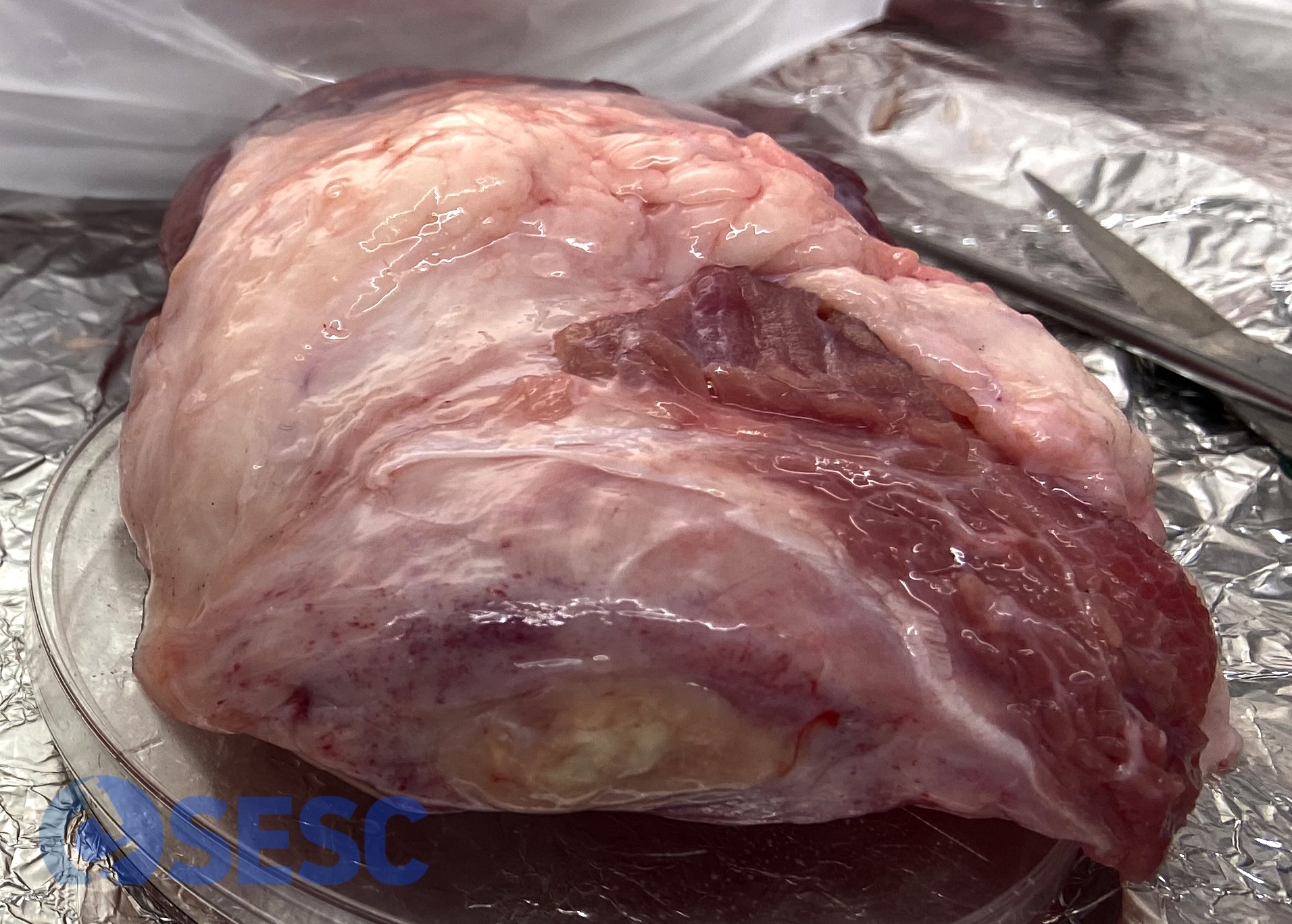
Muscle with lesions from 10 animals were submitted. Although variably, all of them presented similar lesions, ranging from 1 to 4 centimetres in diameter. They presented an indurated consistency, and featured a yellowish central area surrounded by a thick rim of pinkish tissue (fibrous tissue). Histopathological study was performed, which revealed chronic inflammatory and necrotizing lesions within the muscle and adipose panicle (myositis and panniculitis), with a prominent lymphoplasmacytic component (with pseudofollicular arrangement) and presence of eosinophilic amorphous material (presumably injected material).
Although not fully conclusive, the inflammatory component of the lesions could suggest a vaccinal origin, as the local stimulation of the immune system elicits a lymphoplasmacytic reaction much greater than those seen in non-vaccine pharmacological reactions. At the documentation regarding the food-chain information it was recorded that these animals had been vaccinated 3 weeks prior to slaughter. The size, characteristics of the inflammatory infiltrate and chronicity of the lesions are compatible with the injection of a vaccinal product 3 weeks prior to slaughter, as detailed in the carcass documentation. (AC)

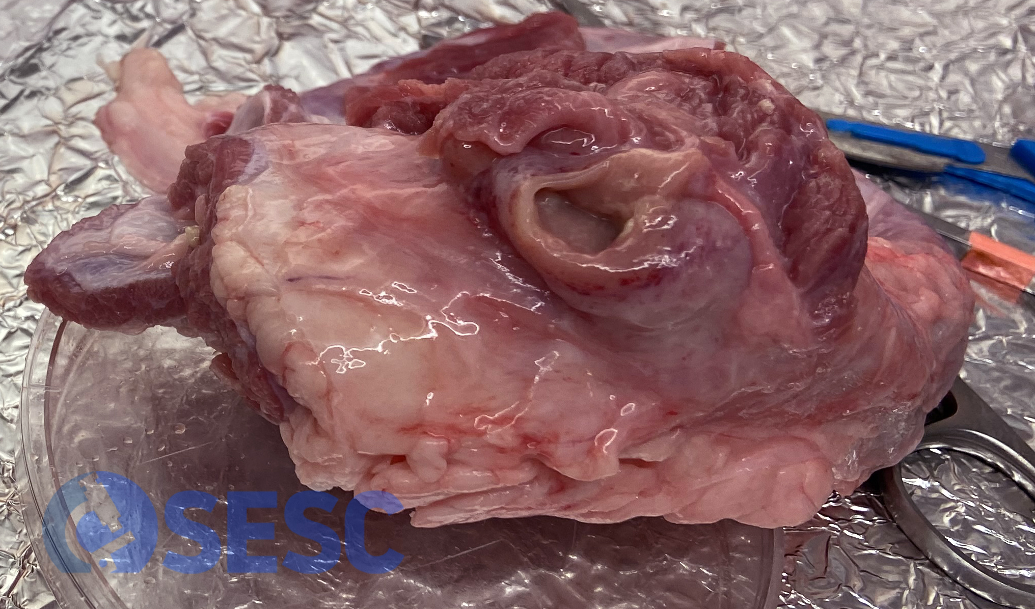
LThe fascia of this muscle was thickened and indurated focally, and at the cut section revealed yellowish material in the central area.
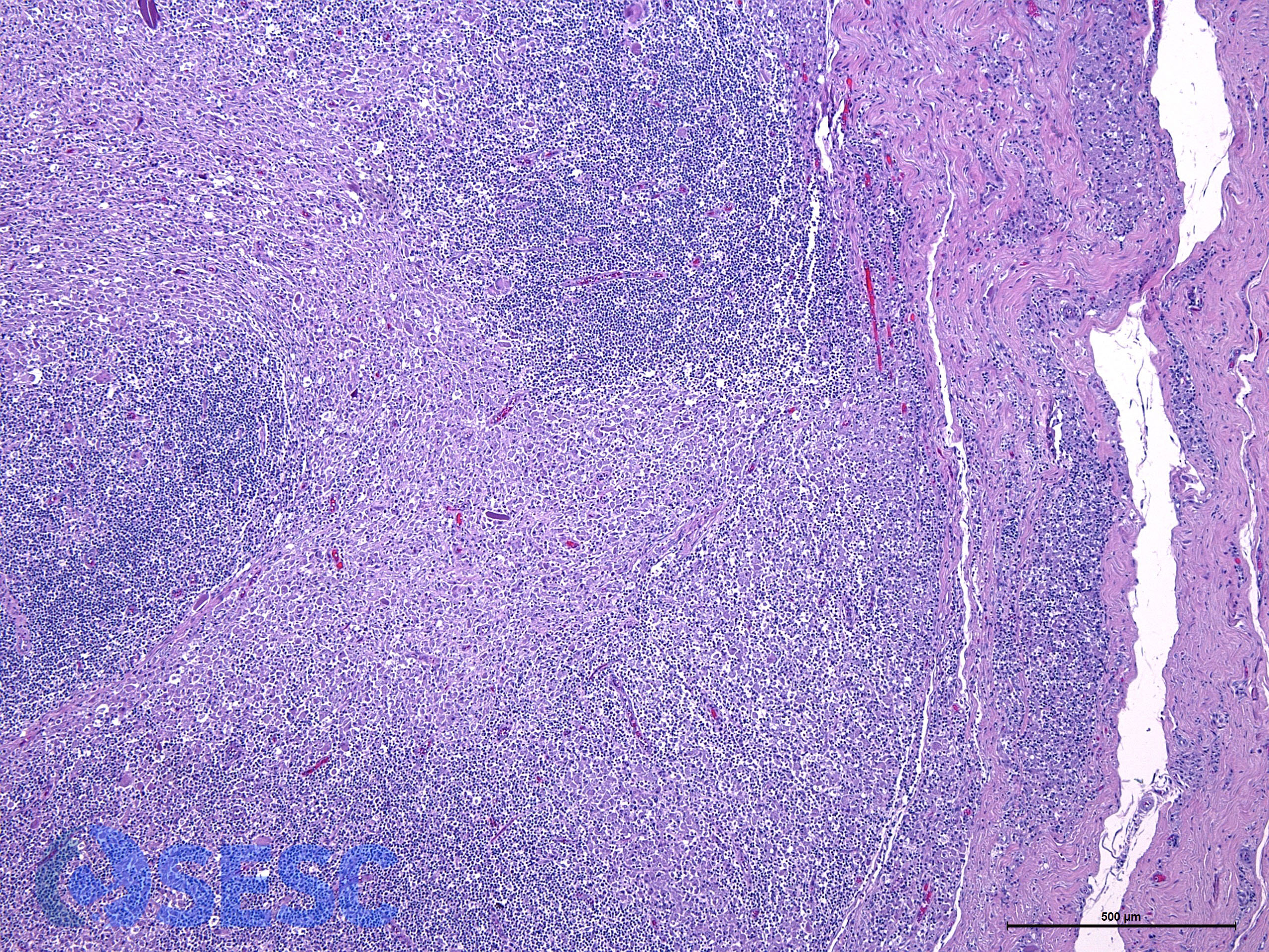
Histologic detail of the lesion. A prominent inflammatory reaction can be observed, mainly lymphoplasmacytic and to a lesser extent, macrophagic, in pseudofollicular disposition.
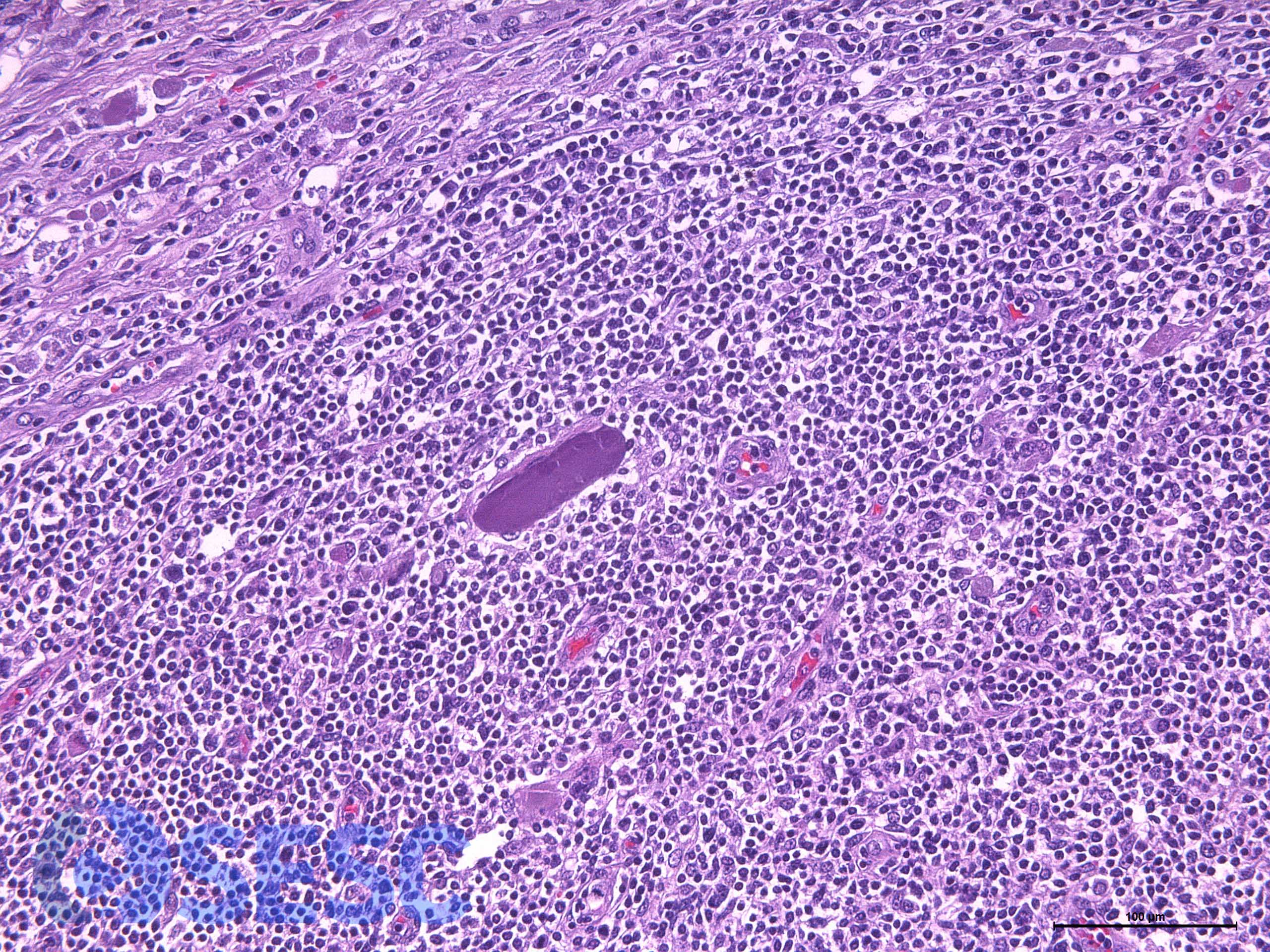
Scattered within the inflammation, accumulation of amorphous eosinophilic material can be seen, presumably injected material.
Similar lesion, in this case affecting mainly the muscle but in a similar manner. In the periphery of the lesion, the fibrosis is progresively replacing the muscular tissue.
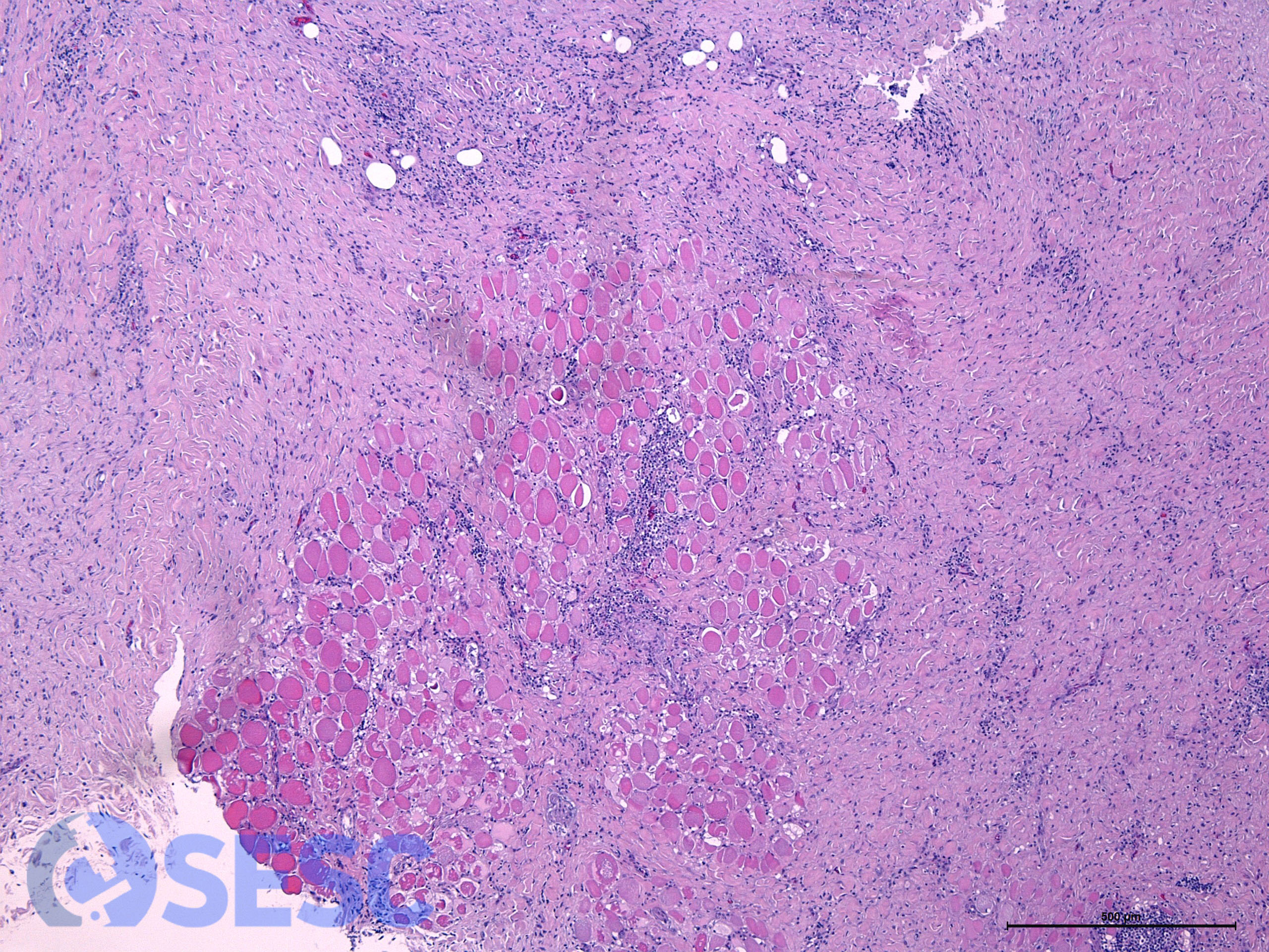
Histologic detail of the periphery of the lesion above. Abundant fibrous tissue and lymphoplasmacytic inflammation can be seen replacing and surrounding the muscular fibers. .
Some of the lesions showed a cavitary appearance, in which the peripheric reaction was less prominent and presented a central cavity with fluid content.
