Lipomatosis cordis in veals
In this post we present two cases of whitish lesions in the heart of calves, both of mixed breed and about 10-11 months old. The histopathological study showed that the lesions consisted of focal areas of well defined and non-encapsulated adipose tissue of normal histological features. No alteration of the muscle fibers surrounding the lesion were observed nor signs of inflammation.
This lesion was classified as focal myocardial lipomatosis thus compatible with a lipomatosis cordis. It is interpreted as a replacement of muscle tissue by adipose tissue due to previous muscle tissue degeneration or necoris, but, in these cases, the etiology of initial muscle lesions could not be determined. These kind of lesions can be observed also in the carcass.

Male, cross breed, 11 months old Calf. Areas of whitish discoloration of the myocardium (arrowheads).
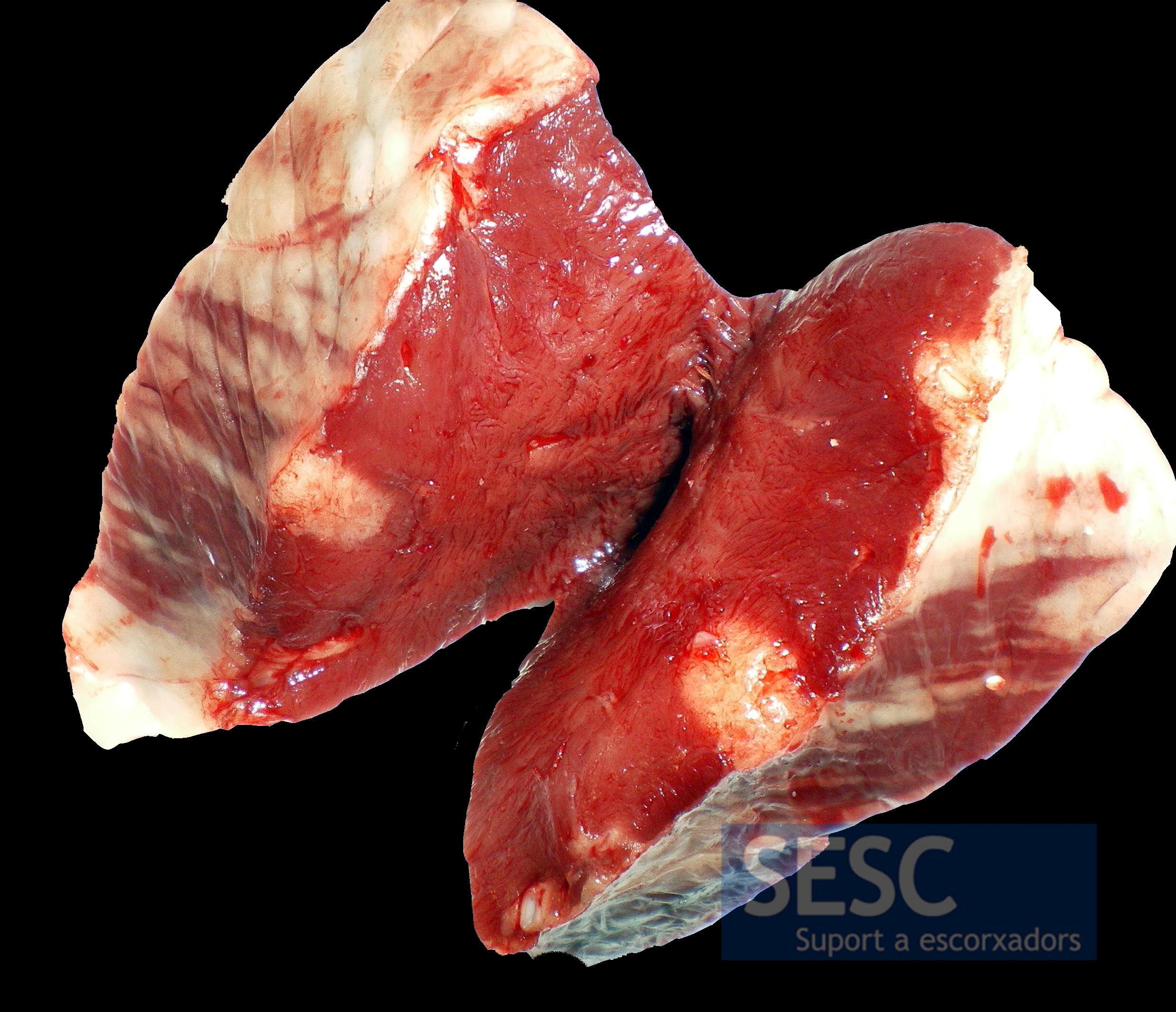
Female, cross breed, 10 months old calf. Two whitish colored nodules in the myocardium.
