17/02/2011
|
Kidney (porcine)
0
Neoplasia in a pig carcass
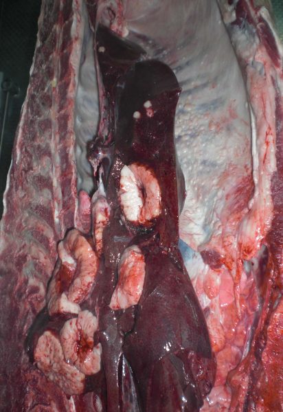
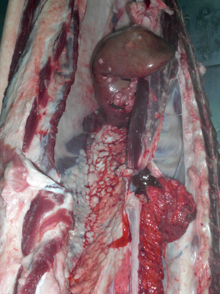
In a carcass of a mixed breed, 3 years old, female pig multiple nodular whitish lesion were observed. The nodules were soft, multifocaly distributed and of variable sizes and were found in the liver, kidneys, peritoneum and lymph nodes.
Histopathological examination of the samples showed that it was a malignant neoplastic proliferation of round cells which was classified as histiocytic sarcoma. It is a low frequency neoplasia in pigs.
The differential diagnosis of these lesions should include, firstly, a lymphoma and other malignancies such as a carcinoma or mesothelioma.


