Nephroblastoma in a pig
Histopathological examination of the mass revealed lesions compatible with a nephroblastoma. It is an embryonic renal neoplasia. In pigs and chickens its the most common primary renal neoplasm. It can be single or multiple and bilateral and can become very large, in pig it rarely metastasizes.
Within the initial differential diagnosis lymphoma was also included as it is one of the most common malignancies seen in pigs and it often involves the kidneys.
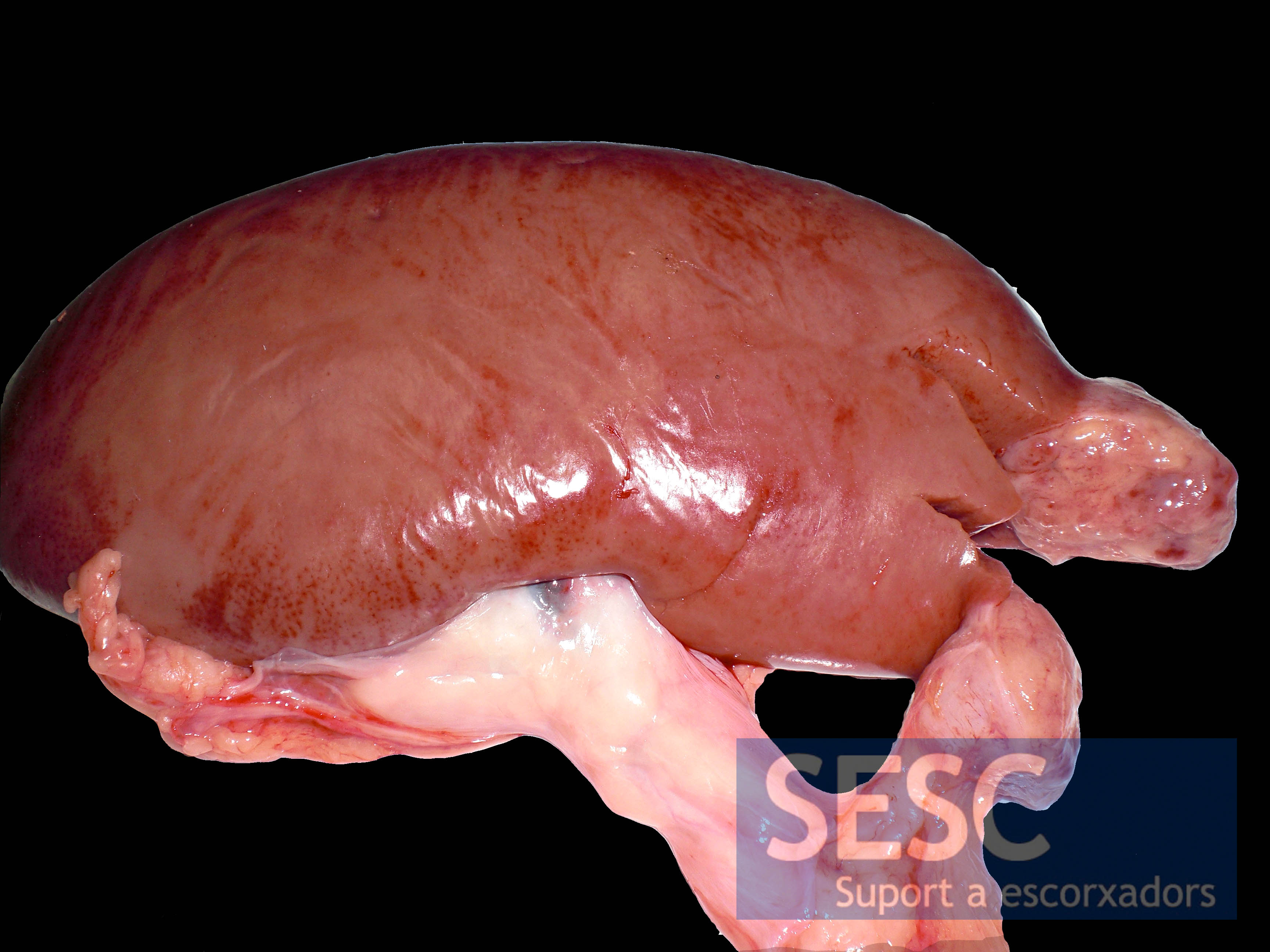
Renal unilateral whitish mass.
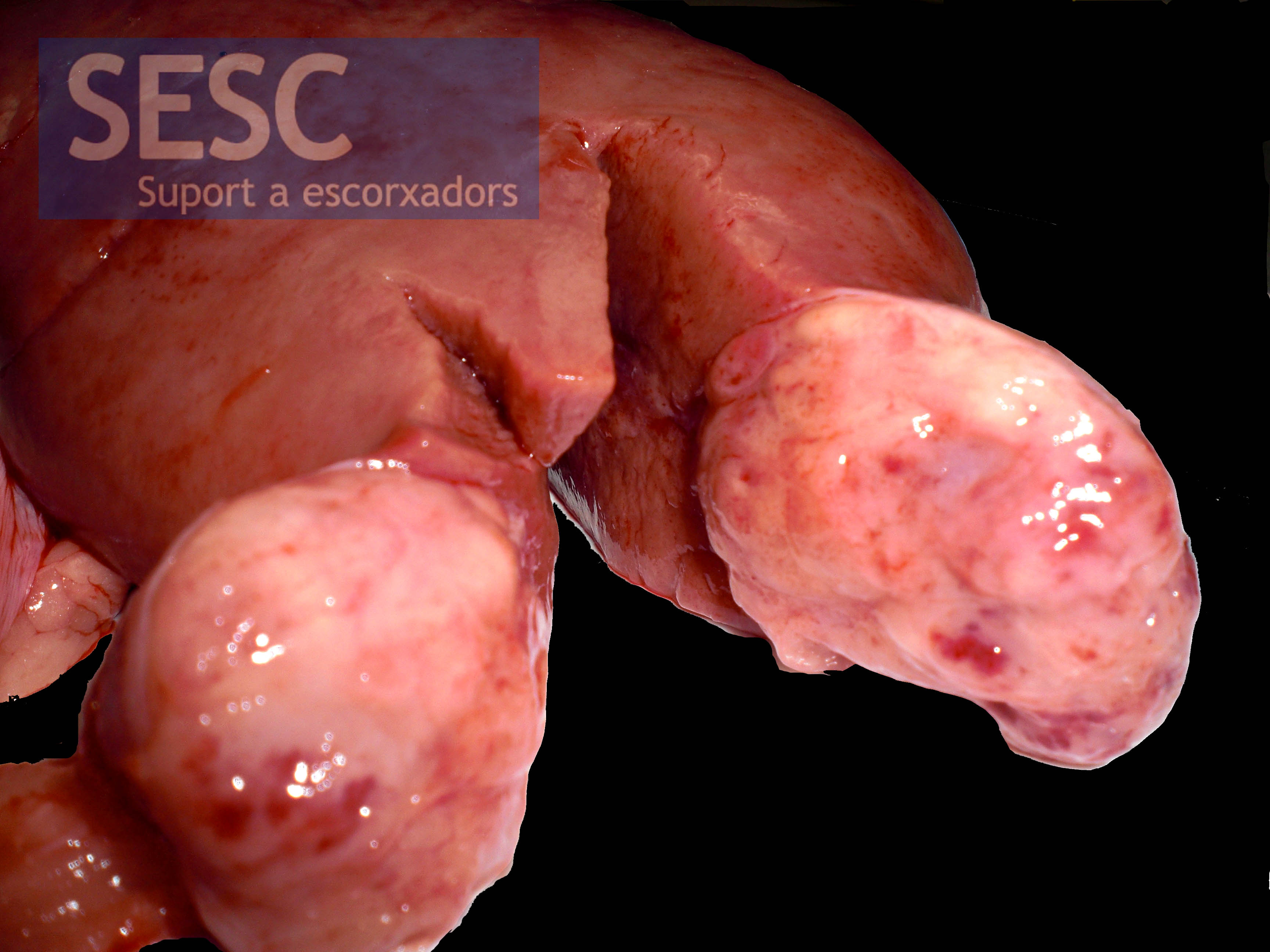
Detail of the cut surface of the mass.

The neoplastic mass was sharply separated from the normal renal parenchyma (asterisk) by fibrous tissue capsule. The growth pattern was solid and densely cellular with formation of multiple primitive epithelial tubular structures.
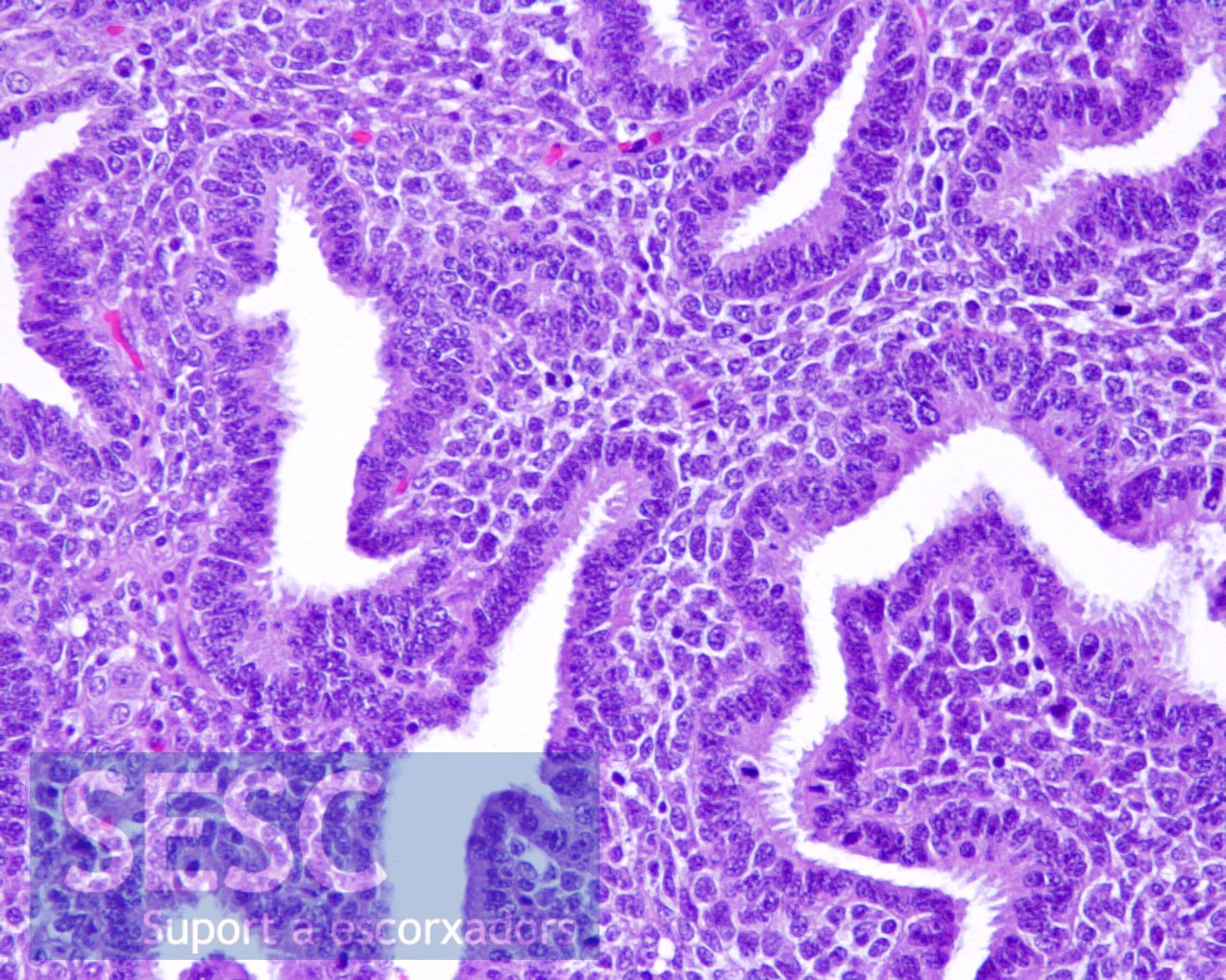
Detail of the primitive renal tubules. In this case differentiation into mesenchymal components which characterizes nephroblastomas was not observed.
