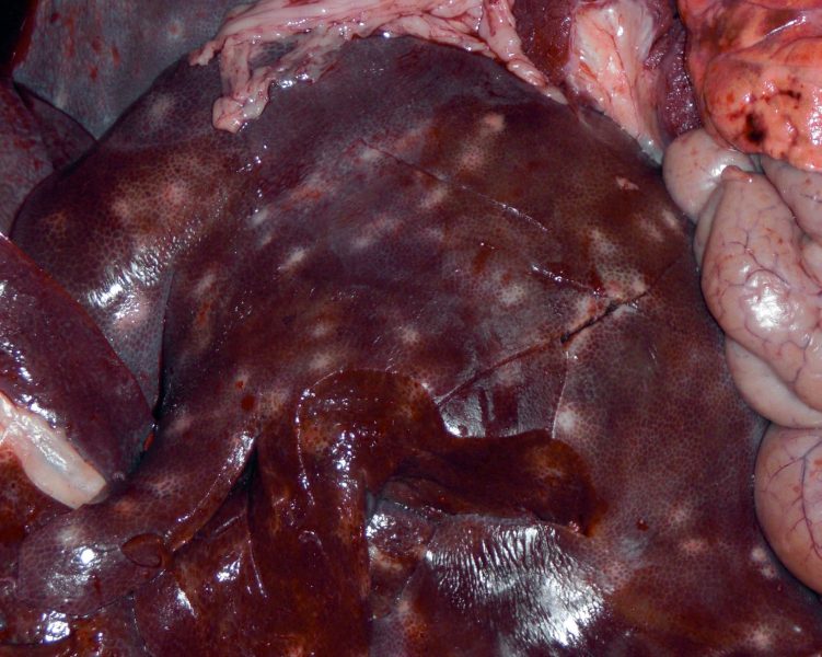
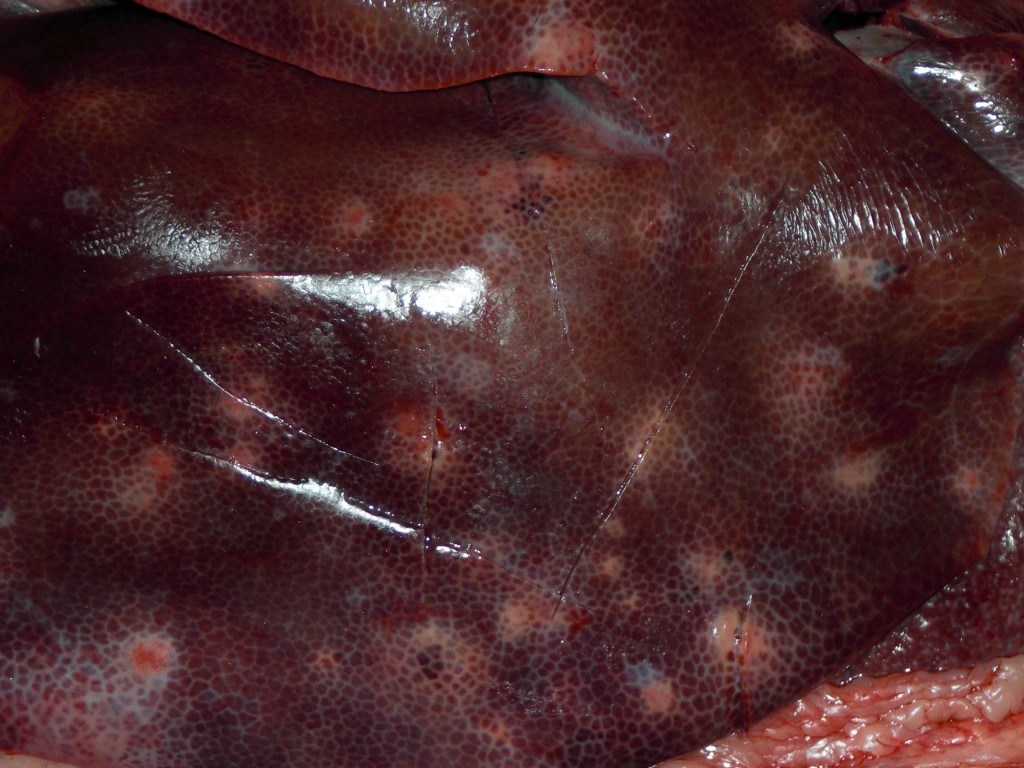
Pig livers with “milk spots”
Liver samples showing white spots were submitted to SESC of two mixed breed 6 months old pig carcasses. Lesions were between 0.5 and 1 cm in diameter, were distributed multifocaly and apparently infiltrated the hepatic parenchyma.
Meat inspectors differential included mycobacteria, Actinomyces spp. and parasites.
The histopathological lesions showed fibrosis accompanied by an inflammatory infiltrate rich in eosinophils and polymorphonuclear leukocytes along with a mild bile duct proliferation.
This lesion is known as "milk spots" in the liver. It is characteristic of a chronic parasitic hepatitis, most likely caused by Ascaris suum. Spots are the result of larvae migration within the liver parenchyma.
Occasionally in earlier stages of the disease, although it was not the case of the samples received, larvae can be observed in the parenchyma, either migrating freely, associated to migration paths or trapped in abscesses or granulomas.
The presence of mycobacteria was ruled out by Ziehl Neelsen staining and PCR.


2 comment(s)
Buenas, se puede consumir la carne de cerdo aunque presente estás manchas el hígado?
Si, el ciclo de Ascaris suum no involucra el tejido muscular. Además, la lesión de manchas de leche es de carácter crónico y ya no contiene parásitos viables.