01/10/2012
|
Head (porcine)
7
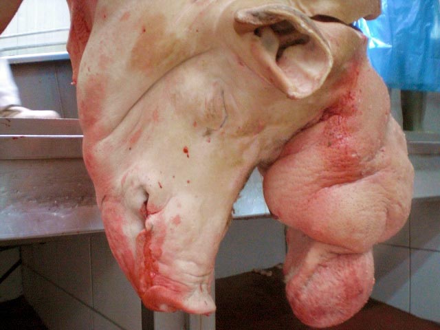
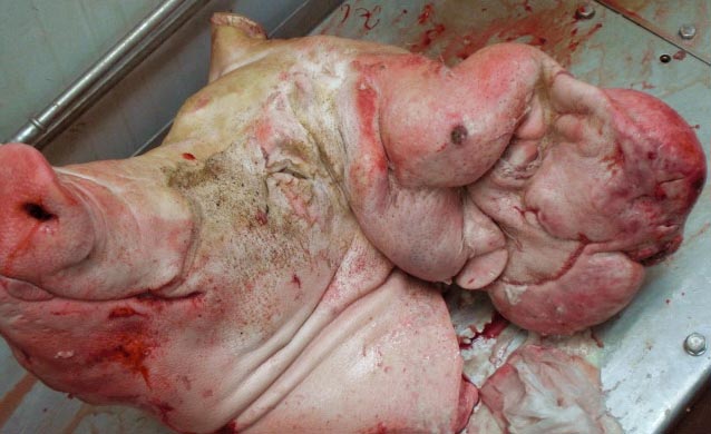
The pig with a giant ear
Inspectors included neoplastic or inflammatory lesions in the differential.
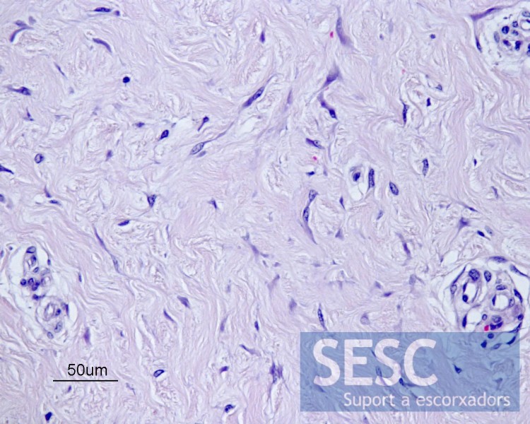
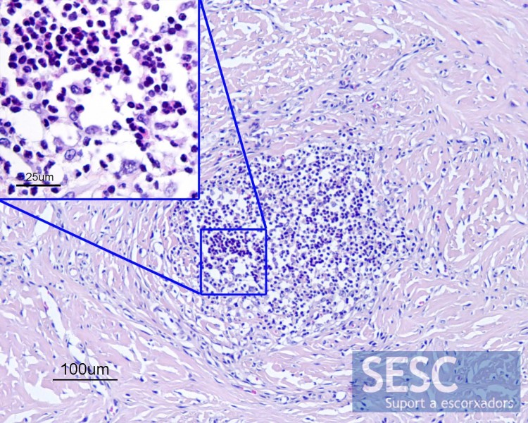
Histopathological examination helped rule out a diagnosis of neoplasia. It consisted of a dense fibrous tissue proliferation, well structured and vascularized along with the presence of a few mononuclear inflammatory cells around blood vessels.
It is a chronic inflammatory reaction most probably caused by trauma. Similar reactions have been described secondary to trampling or piercing the ears to identify animals with an ear tag.


7 comment(s)
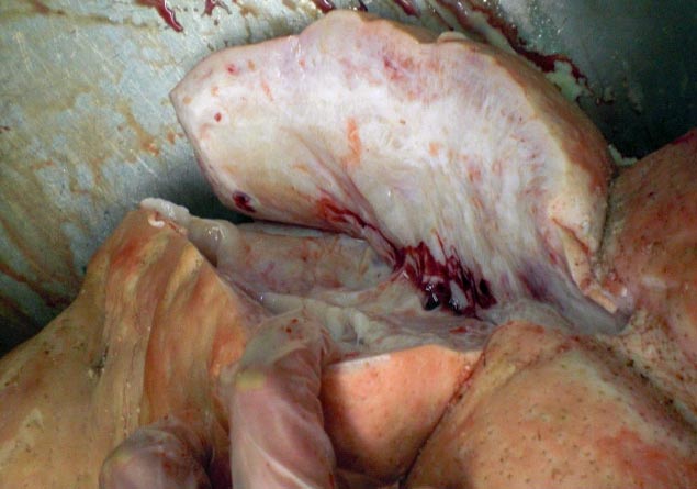
The macroscopic edematous and gelatinous appearance and histopathological characteristics correspond to exuberant granulation tissue.
I am VERY interested in getting high quality images of this to include in a future edition of Zachary’s Pathologic Basis of Veterinary Disease. I have not seen this other than in photos and the ones on this website a great, including the H&E images. Any possibility you could share an H&E recut for future chapter version with inclusion of source in the figure legend?
Thanks,
BNjaa
Thanks for reading our blog Brad! We will contact you through e-mail to send you the files.
Comment from our LInkedIn group:
By Yves Robinson
Veterinary pathologist chez Canadian Food Inspection Agency
Have seen a few similar cases over the years in market hogs. In one of these cases of perichondritis of the ear we found few pyogranulomas with Gram positive cocci. Very interesting.
Thank you for the interesting discussion.
We have added a couple of histopathological images of the lesion. The lesion mainly consisted of mature, well differentiated, connective tissue. No neoplastic proliferation could be identified.
As for the inflammatory findings, scarce and isolated pyogranulomatous foci were identified but no asteroid bodies or Splendore Hoeppli phenomena were observed, nor accumulations of bacteria.
I am pretty sure that this is the right answer: This is a case of auricular elephantiasis caused by Staphylococcus aureus. Histologically pyogranulomatous inflammation (botryomycosis) can be found. The condition is rather common in Danish finisher pigs.
Further reading: Kvist et al. (2002): Evaluation of the pathology, pathogenesis and aetiology of auricular elephantiasis in slaughter pigs. JOURNAL OF VETERINARY MEDICINE SERIES A-PHYSIOLOGY PATHOLOGY CLINICAL MEDICINE Volume: 49 Issue: 10 Pages: 517-522.
A traves de Google reader hemos recibido este comentario:
Saludos, la imagen macroscópica, a mi parecer no sugiere una reacción inflamatoria ni aguda ni crónica, pues en el primer caso habría congestión y el tejido estaría ruborizado, con posibles hemorragias, edema, etc. En el segundo caso (inflamación crónica), si existiese fibrosis, el tejido estaría mas compacto, indurado y no tendría ese aspecto edematoso o mucinoso. Yo, particularmente creo que puede tratarse de una neoplasia mesenquimal, tal como mixoma/mixosarcoma o algún Schwannoma.