Tuberculosis surveillance in slaughterhouses
Tuberculosis is a zoonosis caused by bacteria of the Mycobacterium tuberculosis complex. In Catalonia, in the last 5 years, cases of bovine tuberculosis have decreased considerably due to the program being carried out to eradicate this disease.
In a low prevalence scenario as such, passive surveillance based on detection of lesions compatible with tuberculosis at the slaughterhouse becomes vitally important to detect new outbreaks of the disease and to control them before they spread to other herds or people.
Tuberculosis has characteristic lesions that involve the presence of granulomas, especially in the respiratory lymph nodes (see Figures 1 and 2). It is therefore necessary to make an accurate diagnosis of all granulomatous lesions observed at slaughter. Laboratory analysis (histopathology , PCR , microbiological culture) allow to distinguish whether these lesions are caused by mycobacteria or other etiologies that cause similar macroscopic lesions such as fungi (see Figure 4), parasites, certain neoplasias, etc...
Moreover, tuberculosis does not only affect cattle, goats among other domestic species, are also susceptible and it has been proven that they can transmit the disease to cattle herds. We must also pay attention to the presence of TB lesions in wild reservoirs, especially the wild boar which is a reservoir of tuberculosis in the Iberian Peninsula .
Below are images of some cases arrived at SESC in the recent months.

Figure 1: Lesions compatible with tuberculosis in the mediastinal lymph nodes of a calf.
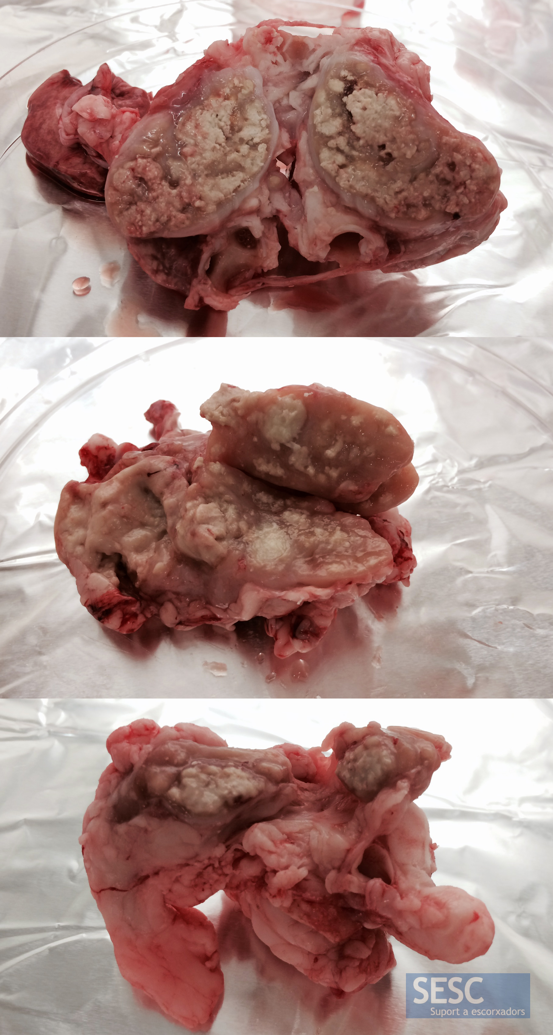
Figure 2; Lesions compatible with tuberculosis in the tracheobronchial lymph nodes of three breeding goats.
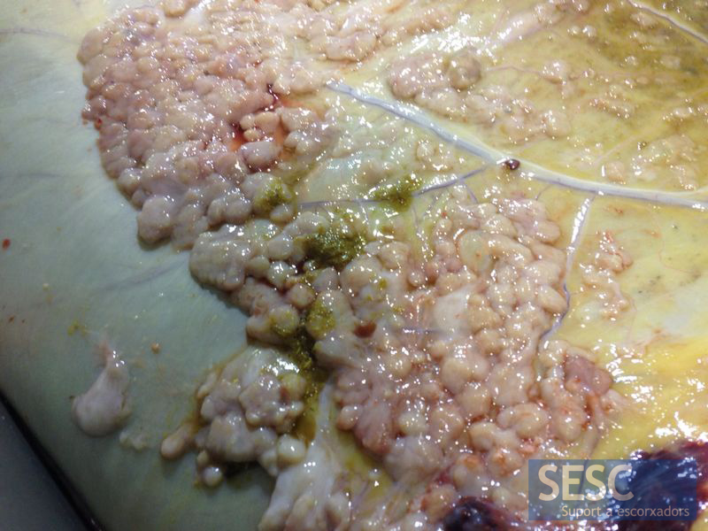
Figure 3: Pearly disease in the rumen of an 8 years old dairy cow.
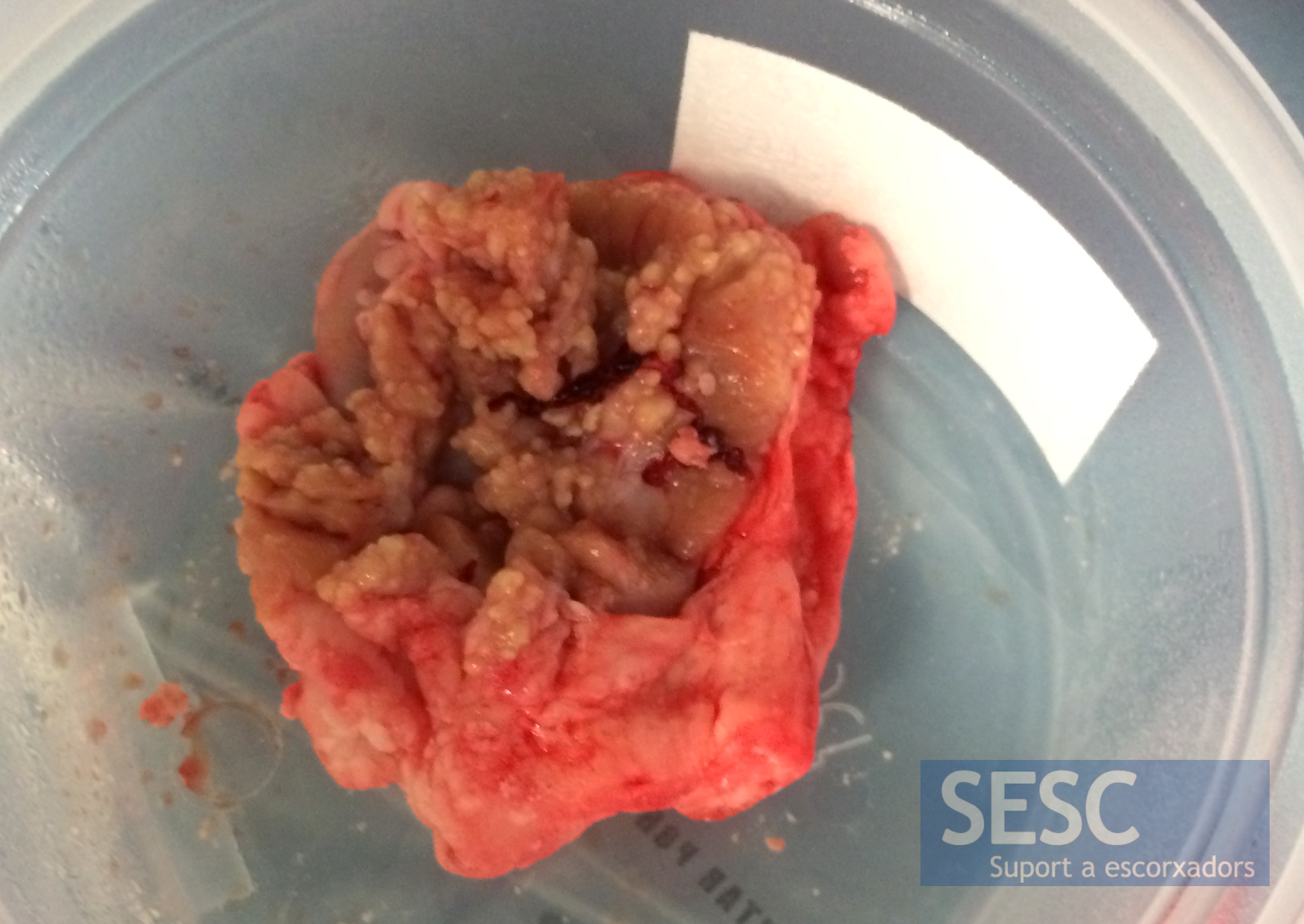
Figure 4: Granulomatous lymphadenitis of the mesenteric lymphnode in a 9 months old calf. The histopathological study showed that the lesions were caused by fungi.

4 comment(s)
It is the best source of information for abattoir surveillance. Can I get the PDF of different working documents? I shall send you mine.
You can find them at the Agriculture department website:
http://agricultura.gencat.cat/ca/ambits/ramaderia/sanitat-animal/
But I’m afraid there’s no english version.
Comment from the “European Animal Health Professionals” group in LinkedIn:
By Giancarlo Belluzzi
Good considerations! But, take care: passive surveillance in the slhouses should be combined with purified proteine derivative interventions and, lastely, with Interferon test. Moreover traditional inspection is frustrated by last EFSA opinions on the biological hazards and risks.
Excellent pictures. Very valuable information for pathologists in USA with little opportunity to see gross lesions of granulomatous inflammation. Well done.