What is your diagnosis? (136)
At the end of the post you can vote for the diagnosis you think is most correct!
Click on the option that you think is correct and press the "Submit" button to find out if you got it right.
We received samples from an 8-month-old cross-breed calf that presented nodular lesions in the lung and lymphadenomegaly of the thoracic lymph nodes. The meat inspectors suspected bovine tuberculosis. (EV)
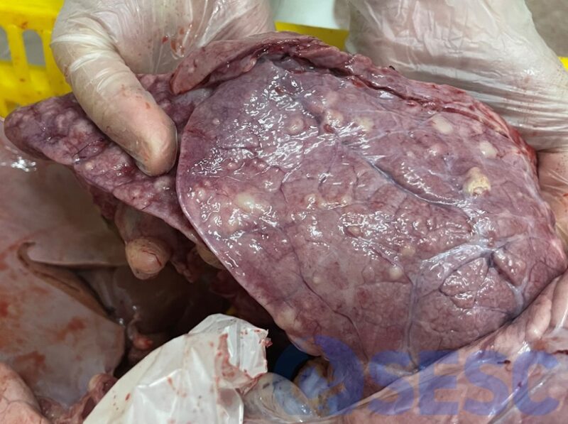
Cattle. Lung. Nodular, prominent, multifocal, generalized lesions of whitish color.

Cattle. Lung. Nodular, prominent, multifocal, generalized lesions of whitish color.
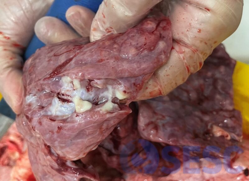
Bovine. Lung. In the section, the presence of a whitish, thick exudate with a purulent appearance can be observed.
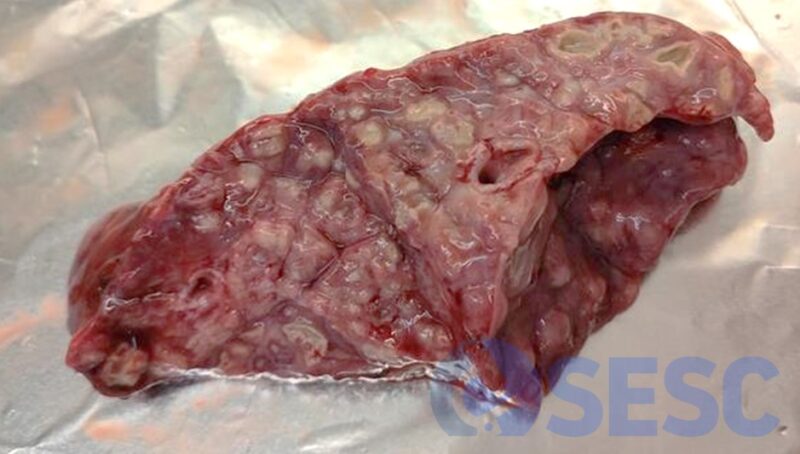
Bovine. Lung. Appearance of the lesion in the sample that arrived at the laboratory.

Bovine. Mediastinal lymph node with increased size and edematous appearance, but without nodular lesions.