Ulcerative dermatitis in a sow
In a 3 year old sow, multiple multifocal, irregular, wet, reddened areas, in the skin of the flanks, abdomen and hind limbs were observed during the ante-mortem inspection. The affected areas had alopecia and whitish vesicles surrounded the ulcers. The post-mortem appearance of the lesions was significantly different and less evident: limited to mild redness and partial loss of the skin of the affected areas was observed.
Histopathological examination of the skin samples taken before scalding of the carcass, evidenced severe chronic ulcerative dermatitis lesions. On the borders of the ulcers hyperplasia and orthokeratotic hyperkeratosis of the epidermis was observed with semiattached crusts. Ulcerated regions had abundant underlying granulation tissue and in the surface, accumulation of bacteria could be seen along with inflammatory infiltrates rich in polymorphonuclear neutrophil leukocytes. In the dermis a perivascular and perianexal lymphoplasmocitic inflammatory infiltrate was seen.
No evidence was found that allowed the identification of the ethilogy of the lesion, in fact, a Grocott staining was used to rule out the presence of fungal structures. Some authors classify this type of lesion, of undetermined etiology, within the Porcine Ulcerative Dermatitis Syndorme or PUDS. The pathogenesis of these lesions is likely to be linked to immune complex deposition.
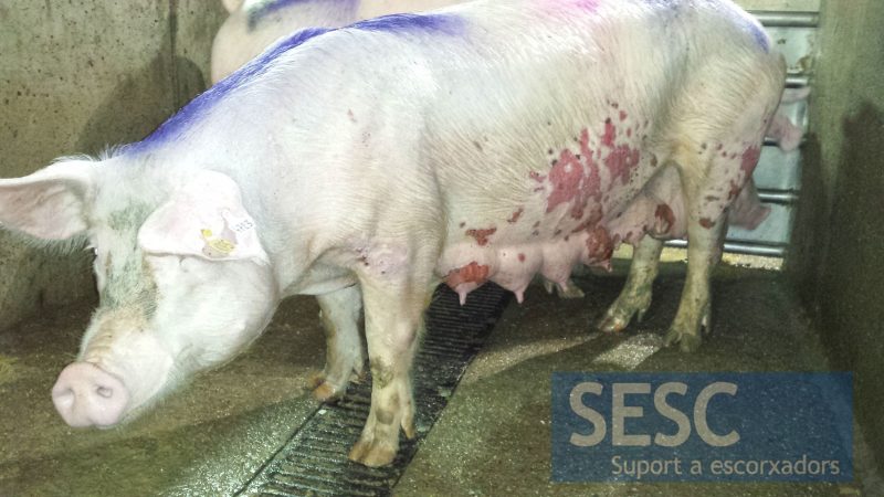
Ante-mortem examination. Multiple ulcers are appreciated affecting the sides and breasts.
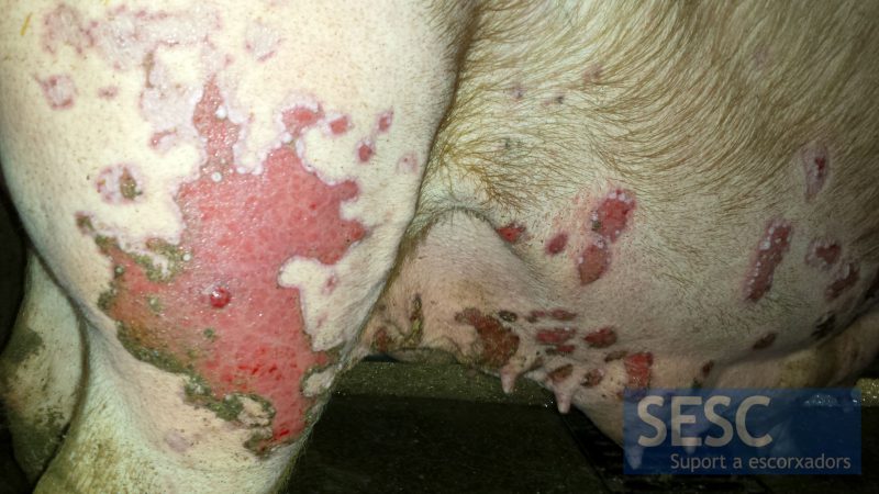
Ante-mortem examination. The skin of the hind limbs was also affected. In the margins of the lesions whitish vesicles were observed.
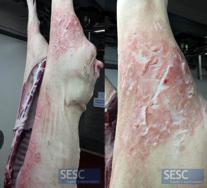
Post-mortem examination. After scalding, the appearance of the lesions has been completely altered and corresponds to red and cracked skin areas.
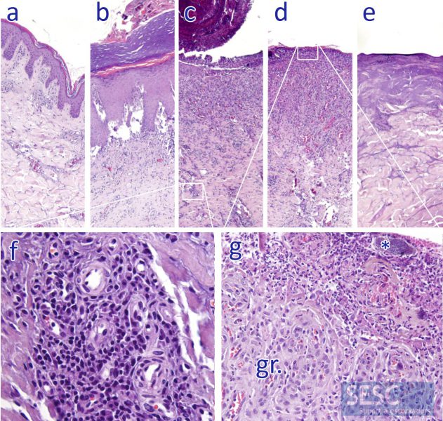
Histopathology. Different portions of the skin were studied. a) Normal-appearing skin. b) Around the lesion, the epidermis was hyperplasied with orthokeratotic hyperkeratosis. c) Beginning of the ulcer with partially detached crust, in the dermis a perivascular lymphoplasmacytic inflammatory infiltrate was seen (expanded in f). d) In the ulcerated areas a layer of granulation tissue (gr. enlarged in g) was appreciated. On the surface, infiltration of neutrophilic polymorphonuclear leukocytes and clusters of bacteria (asterisk) were observed (extended in g). e) Skin sampled after scalding. Coagulation of the structures of the epidermis and superficial dermis prevents the correct assessment of the sample.
