What is your diagnosis? (85)
At the end of the post you can vote for the right diagnosis!
Click on the option that you think is correct and then click on the “Get Results” button to see if you’ve succeeded.
A granulomatous-looking lesion was observed in the caudal mediastinal lymph node of a calf.
So far this year we have analyzed 23 granulomatous lesions in cattle of which 4 have been confirmed as cases of tuberculosis, remember that the annual goal is to analyze 100 granulomas.
Under the microscope, pyogranulomatous lymphadenitis with areas of necrosis, abundant neutrophilic polymorphonuclear leukocytes, and very abundant multinucleated giant cells could be observed.
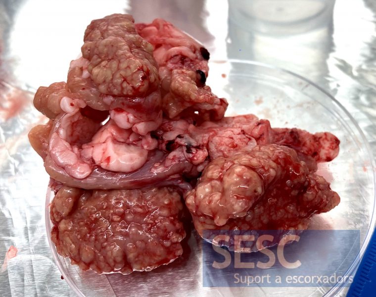
Granulomatous lesion in the caudal mediastinal lymph node of a bovine.
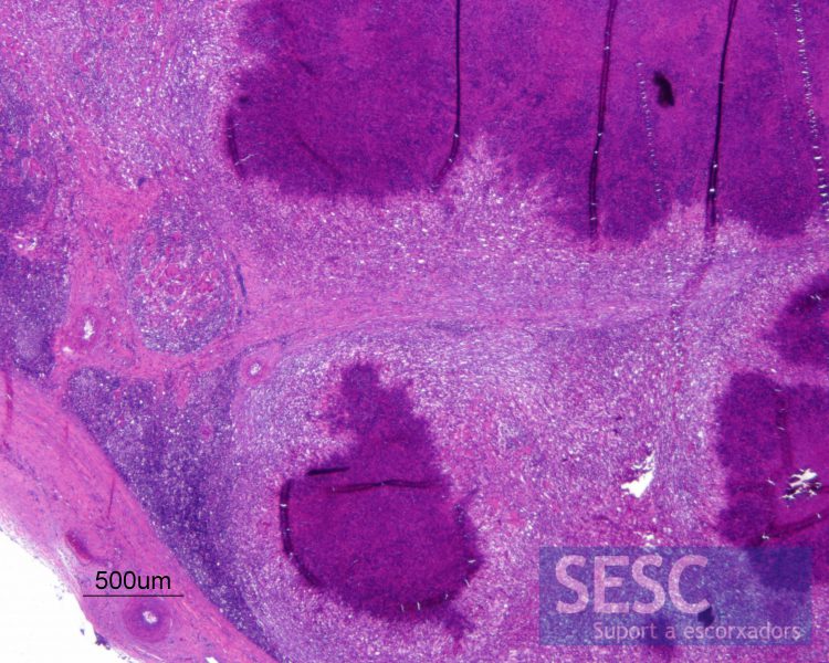
Wide multifocal areas of necrosis (darker areas) are observed under the microscope, with mineralization and abundant pyogranulomatous inflammatory infiltrate.
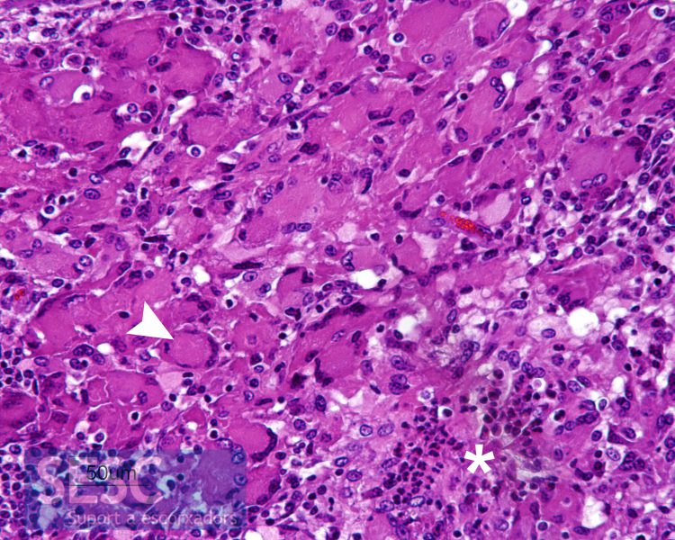
In this enlarged image we can see neutrophilic polymorphonuclear leukocytes (asterisk) as well as very abundant multinucleated giant cells, some of them compatible with Langhans Cells (arrow) that are characterized by presenting the nuclei at the periphery.
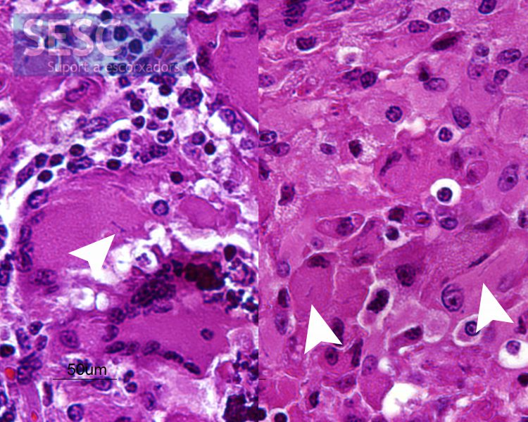
In some of the multinucleated cells basophilic filamentous structures can be observed inside them (arrows).


2 comment(s)
La tinción de Ziehl Neelsen dio un resultado negativo. ¿porqué siendo Nocardia AAR+ no lo evidencia la tinción?
Si bien es cierto que Nocardia puede ser ligeramente acido alcohol resistente, el protocolo normal de tinción de ZN que usamos para detectar micobacterias en nuestro laboratorio no las tiñe.