Chronic liver disease in a calf
In a ten-month-old Friesian calf carcass hepatomegaly was observed along with a hardened consistency and irregular liver surface. Upon section, multiple nodulations were observed. There were no thickened bile ducts or distended gallbladder. The sample was sent as a suspicion of cirrhosis, cholangiohepatitis or necrotizing hepatitis.
Histopathology showed marked interstitial fibrosis in the portal spaces and in the interlobular septa, causing intense disruption of the liver parenchyma, reducing the remaining tissue to multiple regeneration nodules. Among the fibrotic tissue, hyperplasia of the bile ducts was observed, and the presence of individualized hepatocytes that appeared degenerate and atrophic, or swollen with a large nucleus (cariomegaly). Focally, inflammatory aggregates composed of mononuclear cells (lymphocytes, macrophages, and plasma cells) were observed within the fibrotic tissue. It was classified as chronic hepatic fibrosis with regeneration, a lesion that precedes liver cirrhosis.
Hepatic cirrhosis is the end result of continuous and prolonged damage to hepatocytes and/or biliary epithelium and, therefore, it is a nonspecific finding that is observed in chronic phases of different diseases. In ruminants, including cattle, goats and sheep, there are several causes of liver cirrhosis, but the most frequent is chronic liver poisoning. The most common liver toxins are aflatoxin B1 (derived from the fungus Aspergillus spp.) and pyrrolizidine alkaloids (present in plants such as Senecio, Crotalaria and Heliotropium) among others. In all these cases, hepatocytes metabolize said toxins, giving rise to toxic metabolites.
Chronic liver parasite infections (especially Fasciola hepatica) can also cause cirrhosis by affecting the biliary epithelium. In these cases, the lesion results from cholangiohepatitis with fibrosis focused around the bile ducts where the parasites remain.
Jaundice, photosensitization and generalized edema are common consequences in cases of liver cirrhosis and, in some cases, necrosis of other organs such as myocardium. (CL)
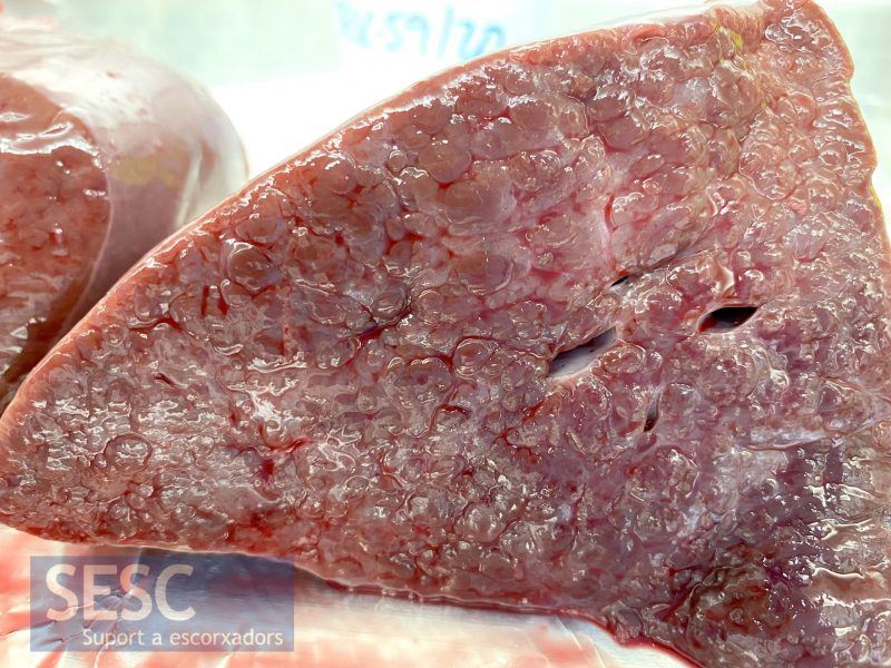
Calf liver. Multiple intraparenchymal prominent nodules of variable diameters. Note the absence of distended bile ducts.
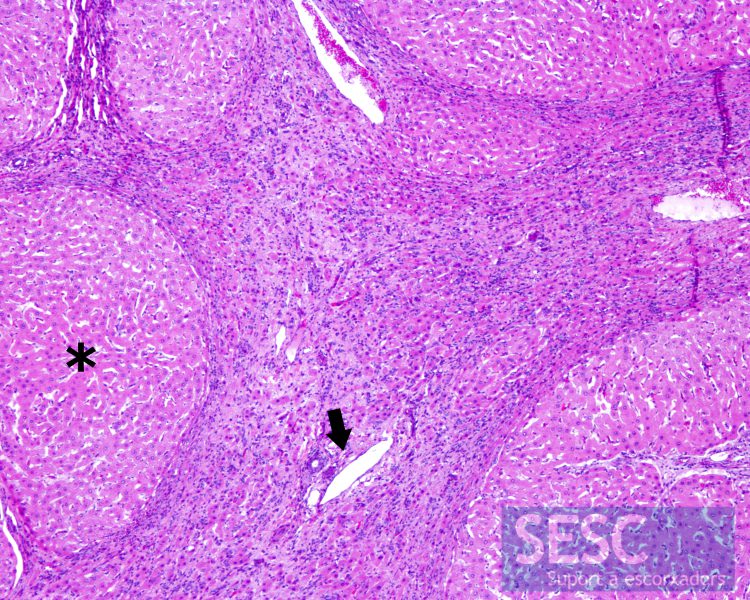
Liver. Hematoxylin and eosin (H&E). Generalized disruption of the hepatic parenchyma, with absence of lobular pattern. Hepatocytes appear aggregated in regeneration nodules (asterisk) separated from each other by a very intense chronic fibrous reaction in portal spaces (arrow) and in interlobular septa.
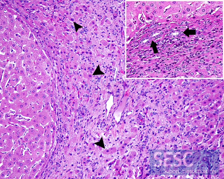
Liver at a higher magnification. H&E. Note the presence of individualized hepatocytes (arrowhead) between the interstitial fibrotic tissue as well as mononuclear inflammatory infiltrate. Inset: bile duct hyperplasia between fibrotic tissue (arrow).

2 comment(s)
Hi! why was the lesion not classified as cirrhosis? The changes seen were not diffuse?
Thanks!
The pathologist that studied the case classified the histopathological lesion as an hepatic fibrosis, they interpret the term cirrhosis as an end stage clinical diagnosis of liver disease (including its clinical consequences) and not a morphological one.