What is your diagnosis? (97)
At the end of the post you can vote for the right diagnosis!
Click on the option that you think is correct and then click on the “Get Results” button to see if you’ve succeeded.
Among the most frequently referred samples to SESC are the granulomatous lesions to rule out tuberculosis. On this occasion we will test your laboratory skills as we show you a special histological staining, would you be able to recognize which technique it is and what are the blue structures that can be observed? (EV)
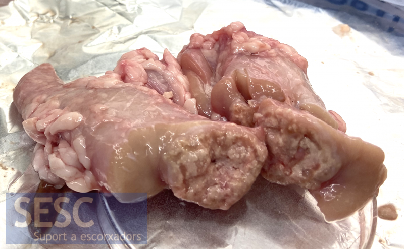
Mineralized granulomatous lesion in the caudal mediastinal lymph node of a 12-month-old cross-bred calf. The animal had a similar lesion in the lung parenchyma.
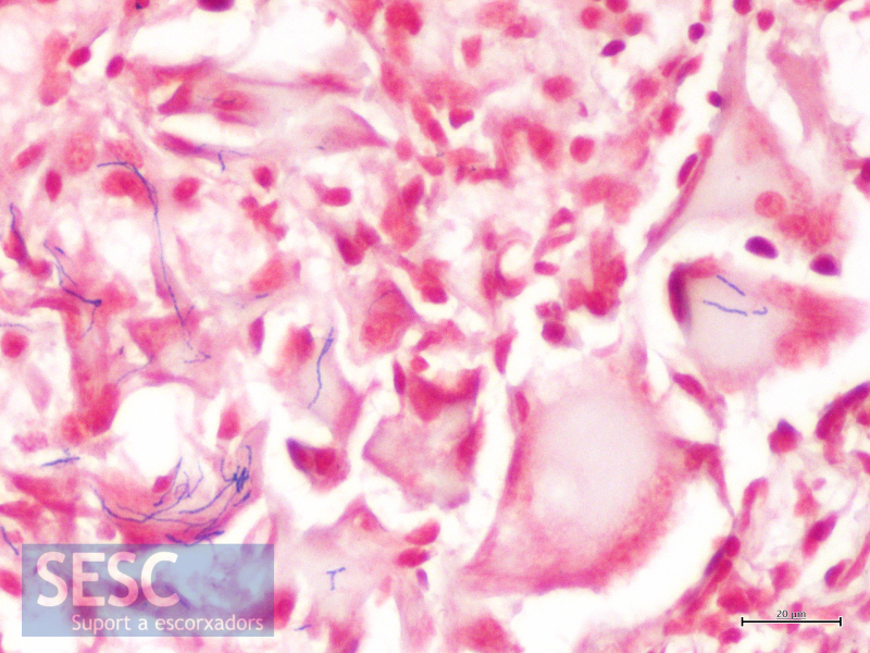
Special staining showing the presence of blue structures stained inside some inflammatory cells. Notice that multinucleated giant cells are observed.
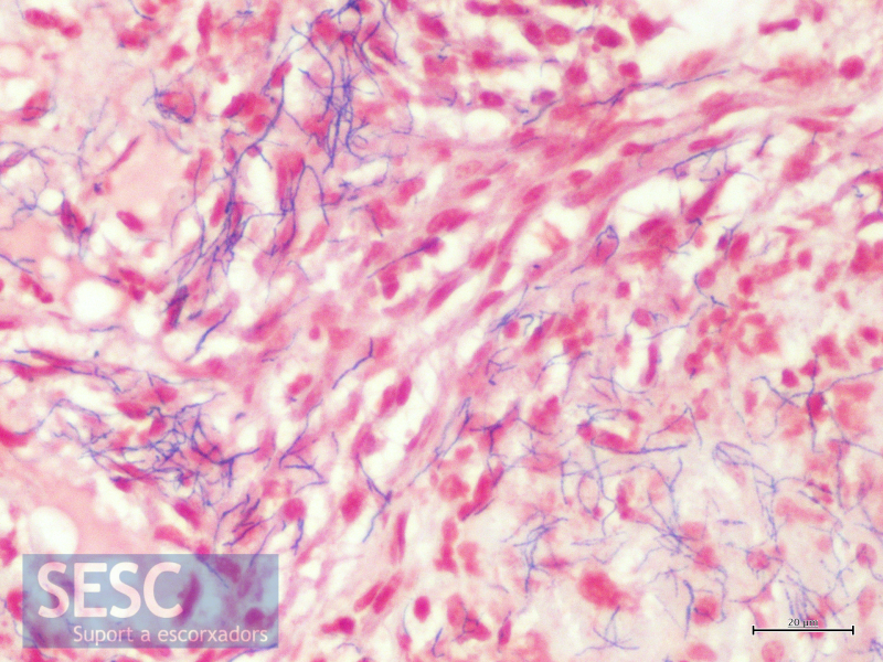
Another fragment of the granuloma where the elongated bluish structures are quite abundant.