E. coli pyelonephritis in a pig carcass
Samples of kidney from a porcine carcass were submitted from a hybrid male, 6 months old. The meat inspectors realized that besides the cortical lesion, showing whitish areas surrounded by a hemorrhagic halo, the renal pelvis was dilated.
After performing histopathology, lesions compatible with a pyelonephritis featuring bacterial presence and suppurative inflammation within renal tubules was seen. A sample was submitted for microbiological culture, from which pure and abundant E. coli colonies were recalled.
Although the appearance of the lesions may resemble an embolic nephritis (such as the one seen in Erysipelas) the distribution of these, near the kidney poles, and the presence of renal pelvis dilation may suggest a potential pyelonephritis. The classical images of pyelonephritis show chronic lesions with abundant fibrosis and cortical loss and retraction, but an acute pyelonephritis habitually shows hemorrhagic and necrotic areas like the ones in this case.
Regarding the common etiology of pyelonephritis, Actinobaculum suis is the classic and specific responsible of these lesions, basically in adult sows, but there are other frequent agents causing the cystitis-pyelonephritis complex, such as bacteria from the genera Streptococcus, Staphylococcus, T. Pyogenes, and the one isolated in the present case: E. coli. (AC)
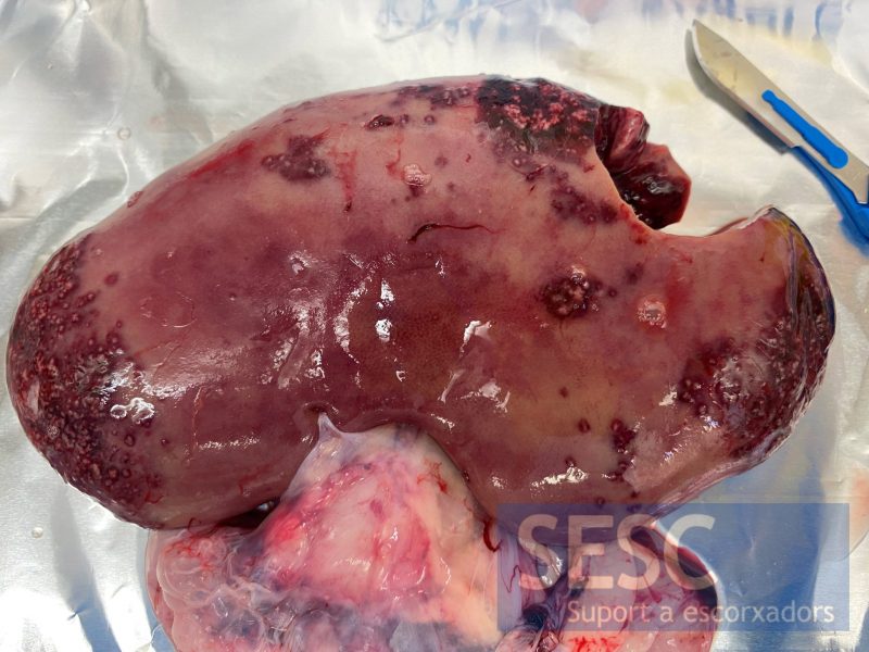
Kidney showing cortical lesions distributed mainly within both renal poles.
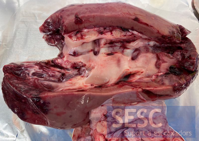
Sectioned kidney. The lesions extended deep through the renal parenchyma, affecting the entire cortex. The pelvis was slightly dilated.
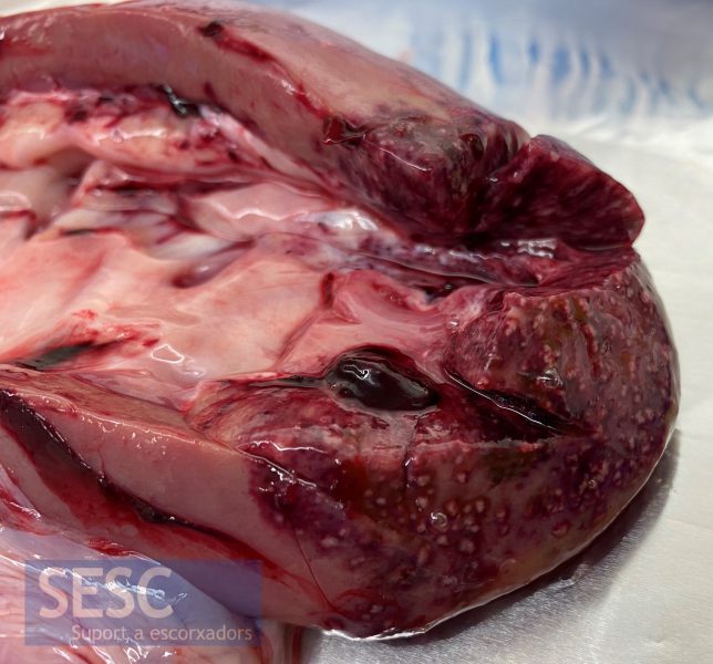
Detail of the cortical lesions. They consist of multifocal to coalescing whitish areas with a reddish halo.
