

White multifocal lesions in rabbit livers
This type of lesion can be associated with different causes, the first two are the most common:
|
CAUSE |
PATHOLOGY |
DIAGNOSTIC METHOD |
| Coccidiosis
(Eimeria Stiedae) |
Hyperplasia and bile ducts fibrosis. | Histopathology: microscopic observation of coccidia.
Stool Analysis. |
| Parasitic migration
(Taenia pisiformis) |
Migration paths.
Granulomas. |
Histopathology |
| Salmonellosis
(Salmonella typhymurium, Salmonella enteritidis) |
Focal hepatic necrosis. | Histopathology
Culture with special enrichment methods. |
| Tyzzer Disease
(Clostridium piliformis) |
Focal hepatic necrosis. | Histopathology
Warthin Starry staining. |
| Listeriosis
(Lysteria monocytogenes) |
Focal hepatic necrosis. | Histopathology
Bacterial culture. |
| Myxomatosis
Poxvirus |
Focal hepatic necrosis. | Histopathology |
| Mycobacterium | Focal hepatic caseous necrosis. | Histopathology Zhiel Neelsen staining. |
| Pseudotuberculosis
(Yersinia pseudotuberculosis) |
Focal hepatic caseous necrosis. | Histopathology
Bacterial culture. |
| Coronavirus | Focal hepatic necrosis. | Histopathology |
| Neoplasia
Lymphoma Uterine carcinoma. |
Histopathology |
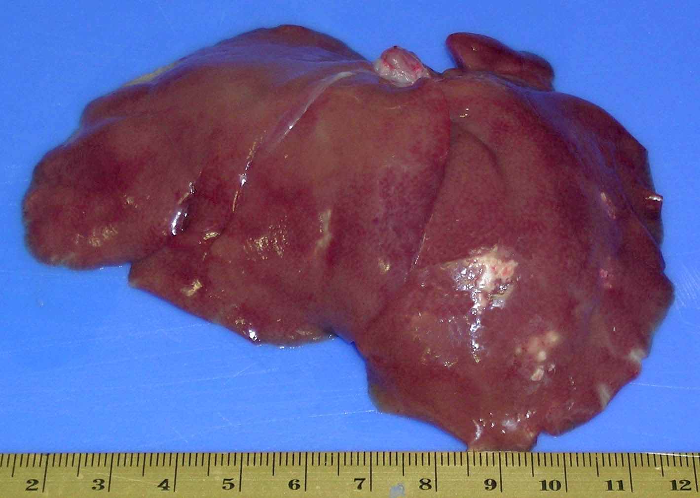
Whitish multifocal lesions. Histopathology could not determine the cause. The microbiological culture was negative, even when pre-enrichment for Salmonella was used. Warthin Starry and Ziehl Neelsen staining did not show the presence of Clostridia nor Mycobacteria. A RFLP study was conducted to detect the presence of pathogenic bacteria witha negative result. The chronic nature of the lesion may prevent the detection of the causative agent.
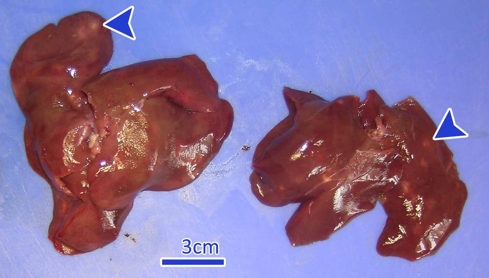
In this occasion the whitish lesions corresponded to hyperplasia and bile ducts fibrosis due to the presence of coccidia (Eimeria stiedae).
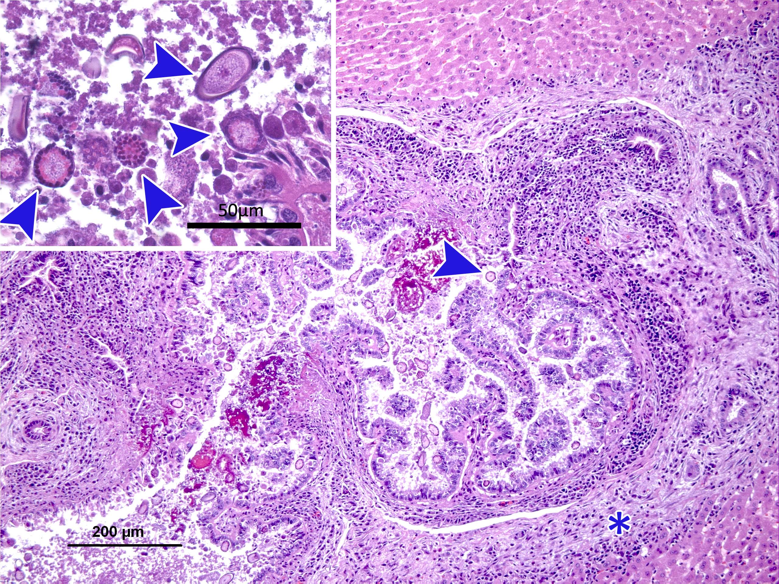
Photomicrograph of an Hematoxylin-eosin staining . Bile duct hyperplasia can be observed as well as periductal inflammation and fibrosis (asterisk) and the presence of protozoal structures inside the ducts and the epithelium (arrows). Top left extended.