Unilateral hydronephrosis and hydroureter in a rabbit carcass
We received an online inquiry regarding the carcass of a two-month-old fattening rabbit that had been declared unfit for consumption. During the ante-mortem examination, no particular abnormalities were detected. However, during the post-mortem examination, upon removal of the viscera, the official veterinary service observed a cystic alteration in the retroperitoneal region, raising suspicion of polycystic kidney disease or similar disorders. Images of the carcass were submitted for evaluation.
The lesions affected only one kidney and the ipsilateral ureter and consisted of a massive dilation of the urinary space, leading to a diagnosis of unilateral hydronephrosis and hydroureter. Hydronephrosis involves dilation of the urinary space of the kidney, specifically the renal pelvis. As a result, the renal tissue (starting with the medulla and then the cortex) undergoes compression-induced atrophy until there is a complete loss of renal tissue, leaving only the capsule, which gives the appearance of a large cyst. In this case, the contralateral kidney and ureter showed no macroscopic alterations.
Possible causes of hydronephrosis include urinary obstructive phenomena, such as urolithiasis occupying the renal pelvis. However, in this case, the alteration must have originated from the distal portion of the ureter, since it is also dilated. The most likely explanation is an ectopic ureter (the ureter does not drain into the urinary bladder) or other developmental abnormalities of the ureter. Interestingly, a complete obstruction involving more distal parts of the urinary tract (such as the urethra) does not cause hydronephrosis, as death due to acute renal failure precedes renal atrophy. The loss of a complete kidney, as in this case, is usually subclinical because the other kidney compensates. In fact, the loss of 75% of renal tissue (equivalent to one and a half kidneys) starts to cause functional impairment of excretory function (AC).

Rabbit carcass. A very pronounced abdominal distension is observed externally.
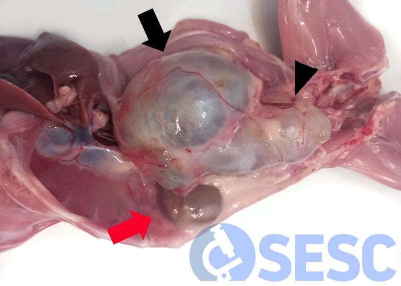
Rabbit carcass. The left kidney (black arrow), as well as the left ureter (arrowhead), are markedly dilated, appearing as a cystic structure filled with urine. The right kidney (red arrow) shows no macroscopic alterations.
