Five reasons a pig carcass might turn red
Since we published the Case of the red pig we have received many inquiries about the origin of reddish lesions on the skin of pig carcasses.
Often these lesions are accompanied by alterations in lymph nodes that may indicate the presence of a systemic process.
We previously reviewed the causes of lymphadenopathy. In this post we will discuss the reasons that can explain the presence of reddish skin lesions and some of the recent cases submitted to SESC.
| 1) Diseases that cause inflammatory lesions (erythematous and/or hemorrhagic) in the skin: |
Each can present particular patterns and location that make them identifiable. If there is haemorrhage in the dermis blood absorption can be observed at the lymph nodes. |
| 2) Causes leading to hemorrhagic diathesis status in the animal | Poisoning by anticoagulants (warfarin, for example, found in rodent poison), thrombocytopenia, vitamin K deficiency, toxicity by copper, zinc, etc ... |
| 3) Traumatic lesions | As a result of fights, scratches or bumps during the transport of animals. |
| 4) Stress response | Pigs respond in a peculiar way to stress during handling. Poor handling or excitement can cause hyperemia and hyperacute dermis hemorrhage/reabsorption of blood at the lymph nodes and erythematous lesions on the skin and subcutis, which should be differentiated from lesions associated with septicemia or viremia. |
| 5) Problems in the management of the carcasses | A lack of bleeding before scalding (Figure 2) or incomplete bleeding could cause images of intense congestion (both of the skin and offal), especially in the lower parts of the carcass. But in these cases the lymph nodes should have a normal appearance.
The possibility that an stunned animal is not dead upon enterning the scaldign tank has to be considered, this would cause a peripheral hyperhemic reaction to the heat. In this case liquid from the scalding tank could be observed within the lungs. |
Additionally, artifacts generated by scalding of the skin makes it virtually impossible to observe the lesions (Figure 5). Therefore, whenever possible, if lesions are observed in the ante mortem examination samples should be taken prior to scalding.
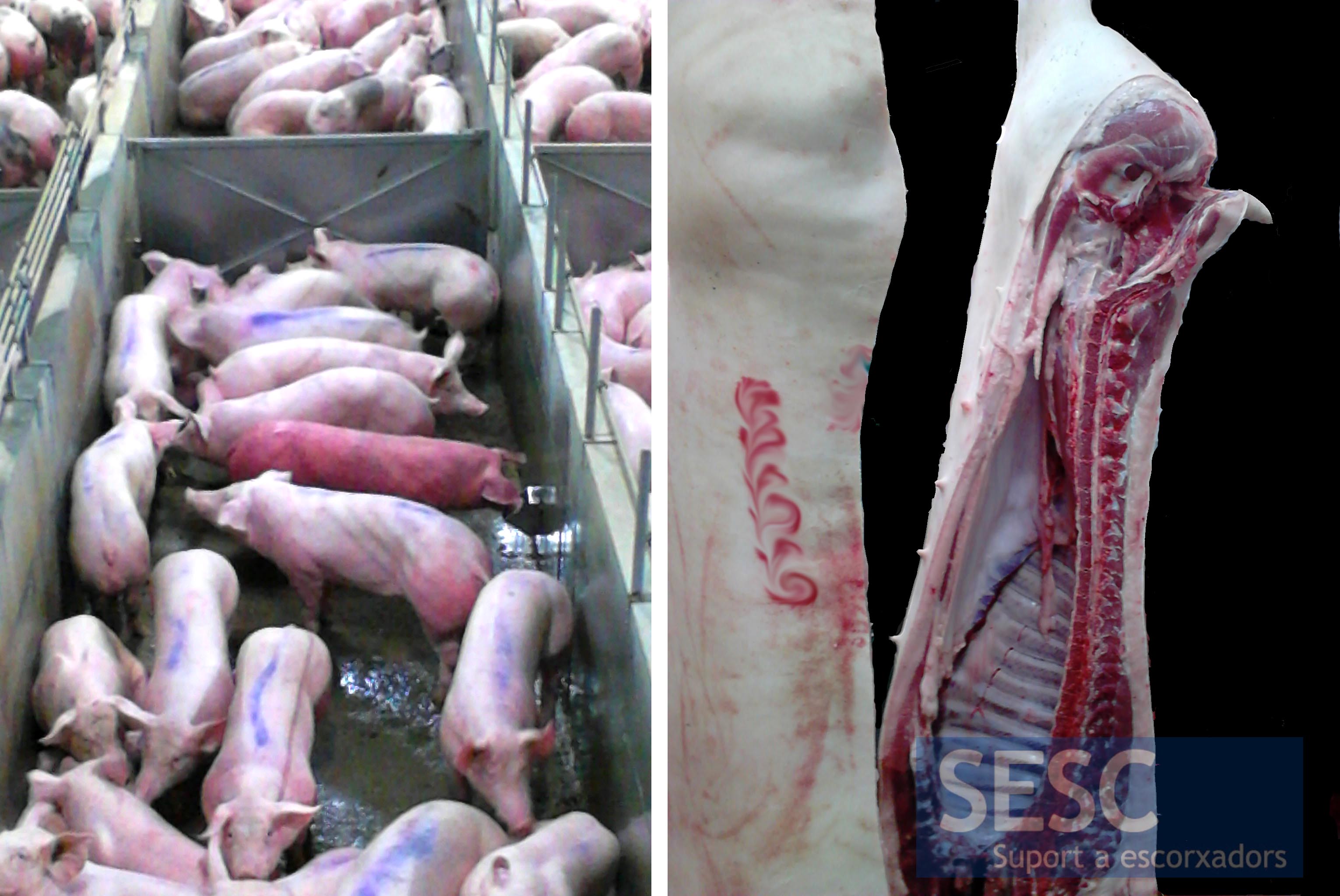
Figure 1. In this case the alteration was detected during ante-mortem examination. In the post-mortem examination, however, showed no pathological changes. It wasn't therefore a true erythema and could be limited to a physiologically active hyperemia and congestion. Possible causes: fights, stress, exercise/strenuous activity, excessive heat or circulatory problems (such as increased blood pressure due to cardiopathy)...

Figure 2: This case corresponds to an animal that was not bled after stunning. It can be observed that gravity pulls down the blood which accumulates in the anterior half of the carcass. A. Image taken 30 minutes after slaughter. B. During gutting we can see that the viscera are more reddened than the rest of the channels. C. In the post-mortem inspection point, ~ 45 minutes after slaughter. The accumulation of blood gives a reddish colouration to the skin of the anterior half of the carcass.
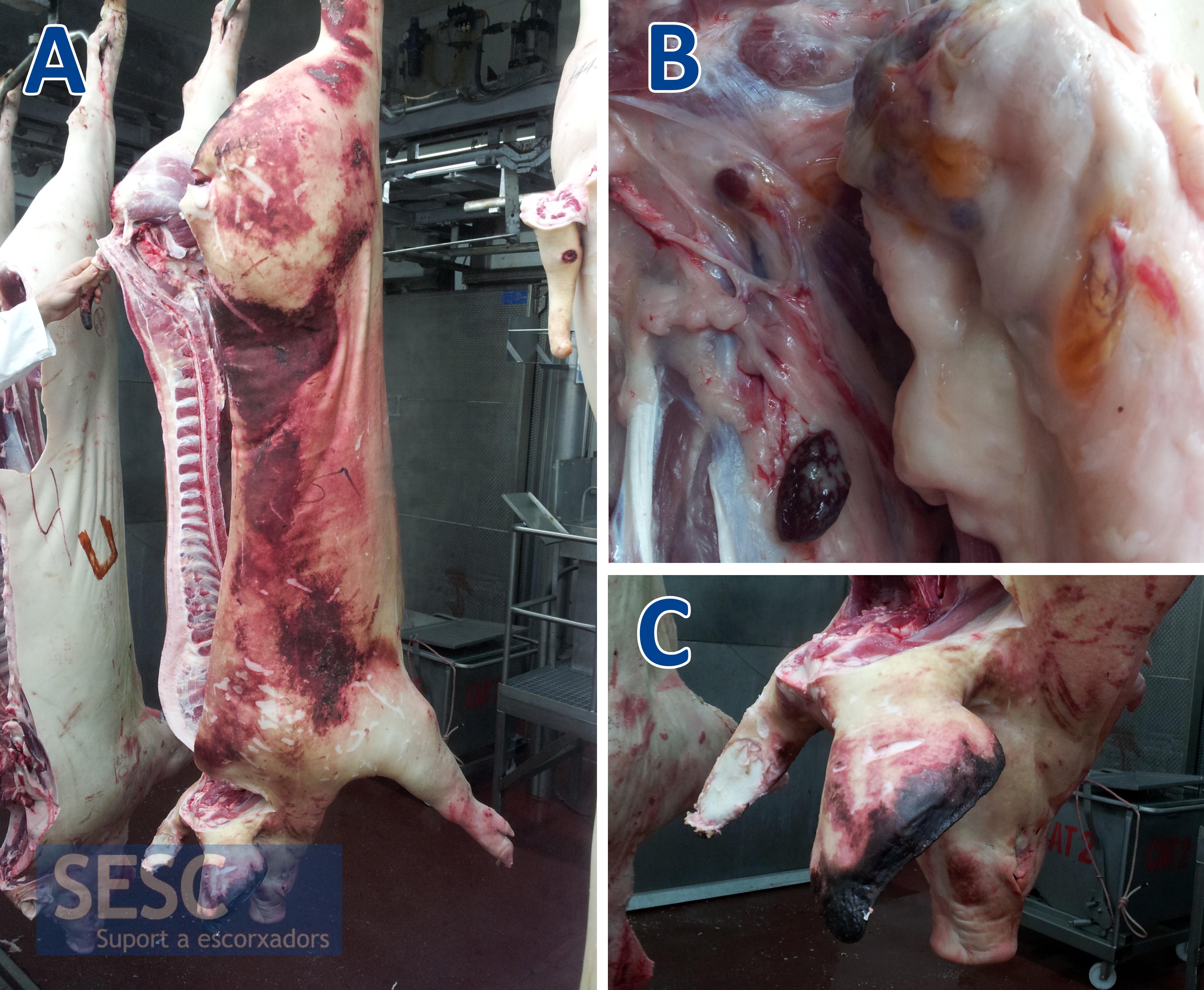
Figure 3. The histopathology of this case determined that the redness of the lymph nodes was due to reabsorption of extravasated blood and therefore result of processes that have caused bleeding in the living animal. It was ruled out that the lymph nodes had lesions of diseases such as PPA or PPC, among others, which may lead to bleeding and necrosis of follicular lymph nodes. In that case, lesions should be observed in larger number of carcasses.
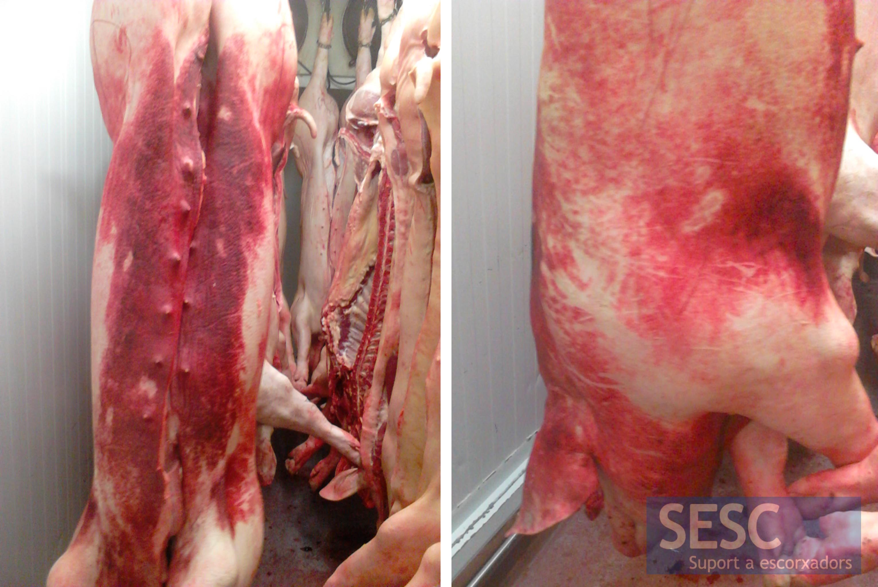
Figure 4: In this case the histopathological examination disclosed haemorrhages on the dermis and therefore was indicative of a pathological or traumatic process. The heart, microscopically showed no abnormalities, grossly it had left ventricular hypertrophy. This could be caused by an insufficient venous return and could also explain the congestion obsereved in the liver and, ultimately, a greater suscetibility to skin hyperemia. There were no injuries on the viscera that indicated the presence of other possible etiologies (viruses, bacteria...). As always, coagulation artifacts of the dermis due to the scalding did not allow to rule out processes that affect the superficial layers of the skin.
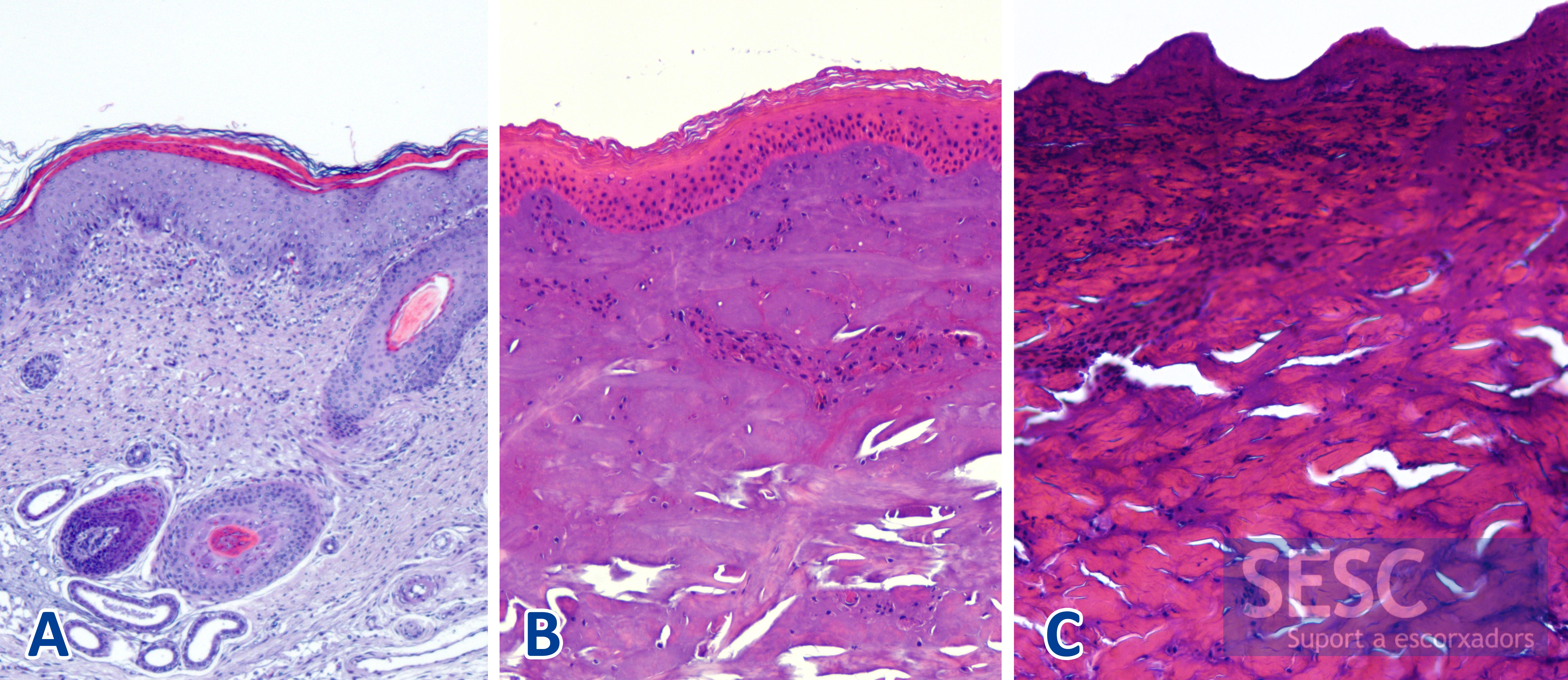
Figure 5: Histological image of the skin of pig that displays artifacts caused by scalding. A. Skin processed before scalding where one can clearly distinguish all skin structures. B and C: Different degrees of artifact due to the scalding of the skin where you can see coagulation of the cutaneous structures that prevents a proper assessment of the sample.

2 comment(s)
A sixth cause has been suggested to us: cardiopathy.
As indicated in Figure 4, an insufficient venous return might predispode an animal to dermal congestion. This cause is in accordance with the appearance of isolated carcassses with this problem.
Comment form our group in LinkedIn:
Claudiu Gabriel Teodorescu
Swine Veterinarian at Garth Partnership. Leading in Pig Health Management.
I have seen recently some carcasses with reddened skin. Those pigs been in the abattoir lairage over the night (more than 12 hours).
I think because they were over crowded and long contact with urine.