Hepatic granulomas in lamb livers
In both cases the lesions were superficial and had adhesions to other serous membranes. Histologically granulomatous inflammatory changes were observed: extensive central necrosis surrounded by abundant epitheloid macrophages, some multinucleate giant cells and presence of eosinophilic polymorphonuclear leukocytes.
The lesions are suggestive of a parasitic etiology and its location is compatible with a degenerate Cysticercus tenuicollis cyst . In case 2 the presence of mycobacteria was ruled out using Ziehl Neelsen, PCR and mycobacteria culture since the samples was submitted as suspected TB case, a disease to which sheep are also susceptible.
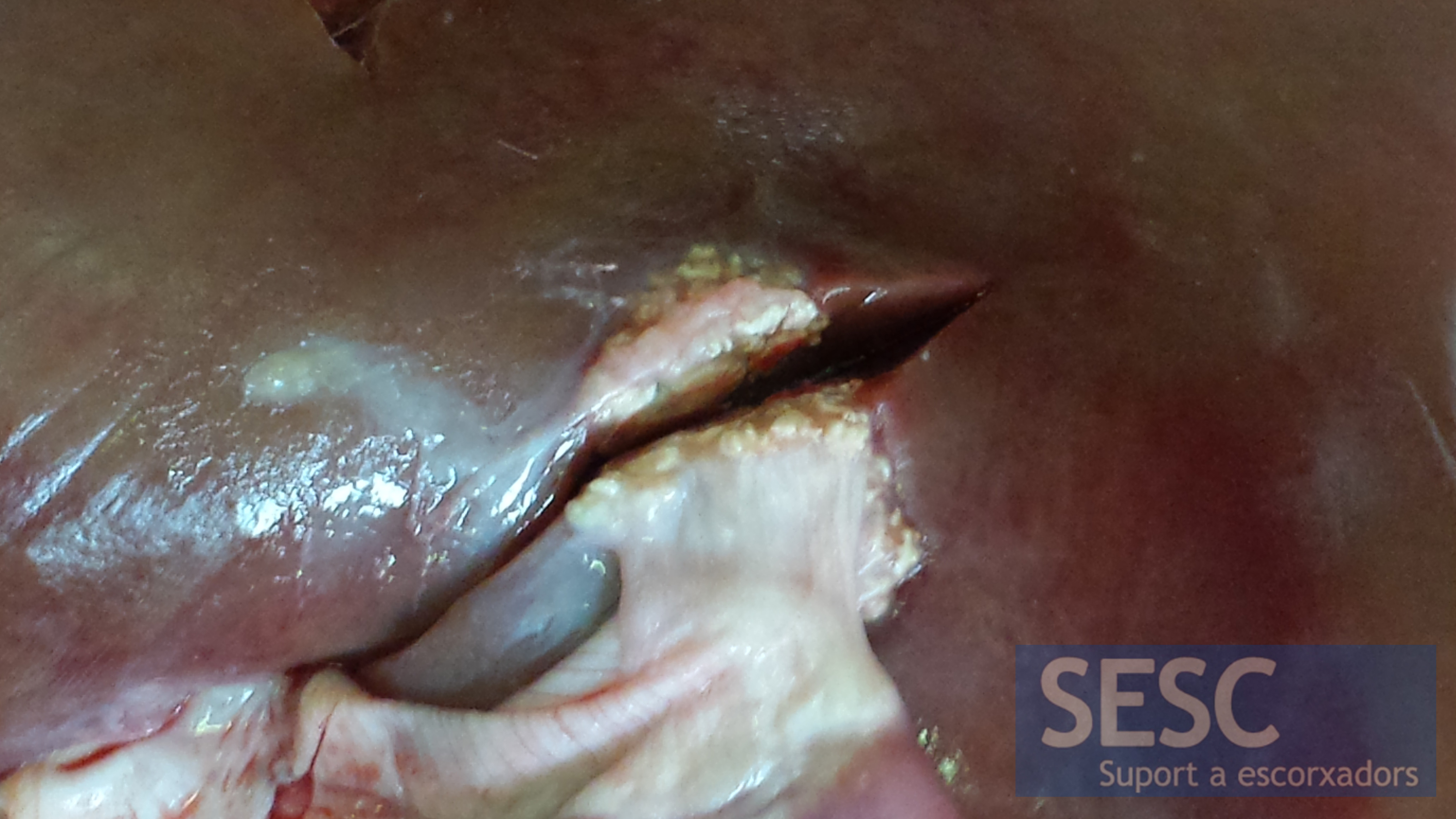
Case 1: Granulomatous lesion in the liver of a three months old lamb.

Case 2: very similar, superficial, granulomatous lesion in another lambs' liver.

2 comment(s)
Comment from Veterinary Pathology group in Linked IN:
By Hala el Miniawy
Vice dean at faculty of veterinary medicine
I think its a pseudo-tuberculosis ovis
Comment from Veterinary Pathology group in Linked IN:
Dave Getzy
Pathologist at IDEXX Laboratories
Hi,
I really appreciate you putting these great cases out for viewing. When I was at the Colorado State University Diagnostic Lab, we used to see these lesions sporadically in lambs, with the same histopathology as you describe. Tuberculosis is always a differential, the eos are usually a good clue, but always a good idea to at least do an acid fast stain to be sure. As much as we like to think one animal- one disease, occasionally, especially in some of the more poorer feedlots, more than one disease may be present at the same time.
Best regards,
Dr. Dave G.
Fort Collins, CO