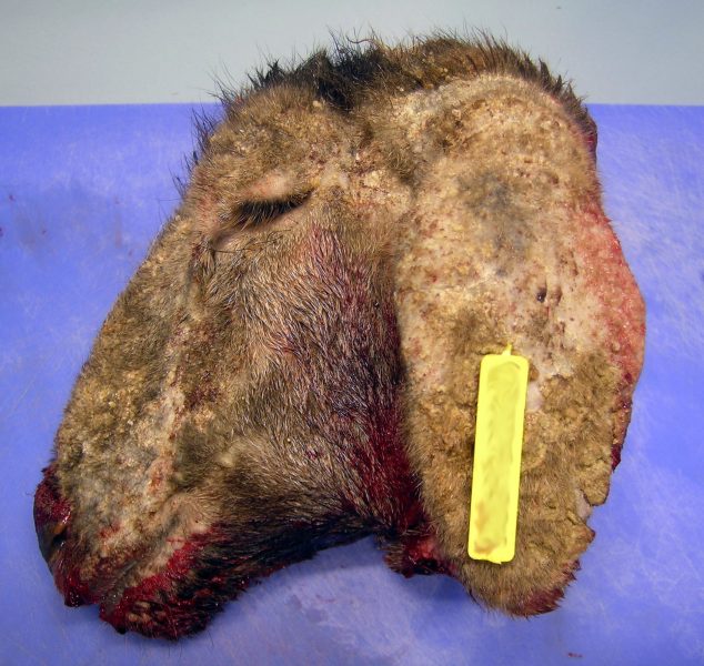
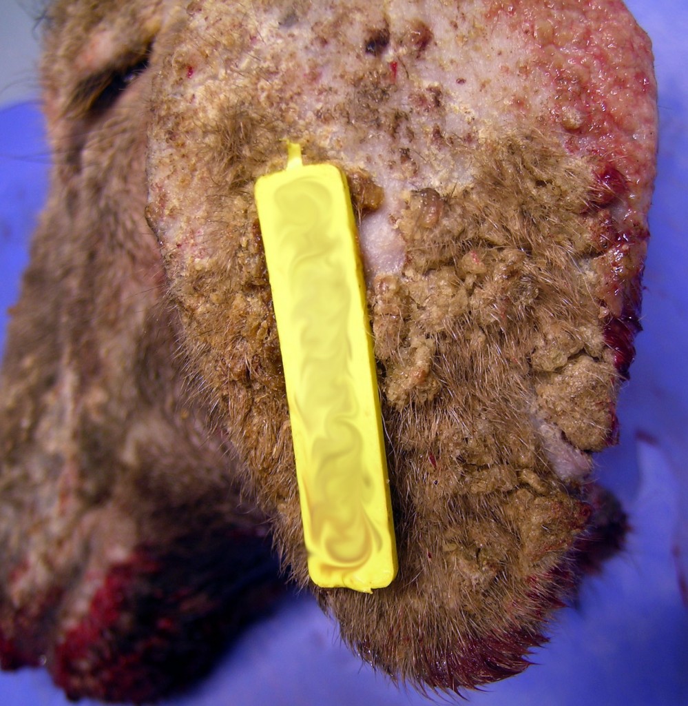
Skin lesions on the head of lambs
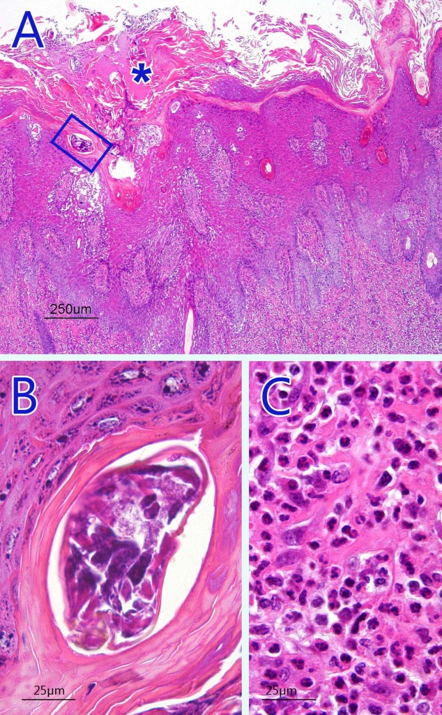
Microscopically a large crust was observed with marked hyperkeratosis and with abundant presence of mites in the stratum corneum. This was accompanied by severe degenerative and inflammatory phenomena of the epidermis with intracorneal pustules. In the dermis there was an intense and diffuse inflammatory infiltrate constituted by numerous polymorphonuclear leukocytes and also eosinophils evidencing a hypersensitivity reaction to the parasite.
The location of the mites and the distribution of the lesions in areas without wool is typical of Sarcoptes scabiei, the causative agent of sarcoptic mange which also affects humans, i.e. it is a zoonosis.

A photomicrograph of a hematoxylin-eosin stain where one can observe an intense hyperkeratosis (asterisk) and the presence of a mite in the stratum corneum (blue box, extended in image B). C: Detail of the inflammatory infiltrate, rich in polymorphonuclear eosinophilic leukocytes, observed in the dermis.


2 comment(s)
gracias por este articulo que nos enseña que todos estamos en peligro con estos parasitos, no se salvan ni los animales mas insospechados
[…] diagnòstic diferencial d’aquestes lesions és: ectima contagiós, sarna i verola caprina […]