Green mass on the urinary bladder of a pig carcass
Histopathological examination of the mass revealed inflammation of the serosa (outermost layer of the wall) of the bladder with abundant eosinophilic polymorphonuclear leukocytes, scarce macrophages and the presence of plant structures and abundant clusters of bacteria. The bladder mucosa (innermost layer) was preserved.
The material that is causing the serositis in this animal is, very likely, of digestive origin. Even though the presence of vegetal material in this location is odd, the only logical explanation is that it might be a sequelae of a chronified (or resolved) peritonitis process that included intestinal perforation.

When the carcass was opened a greenish mass was osberved on the urinary bladder.
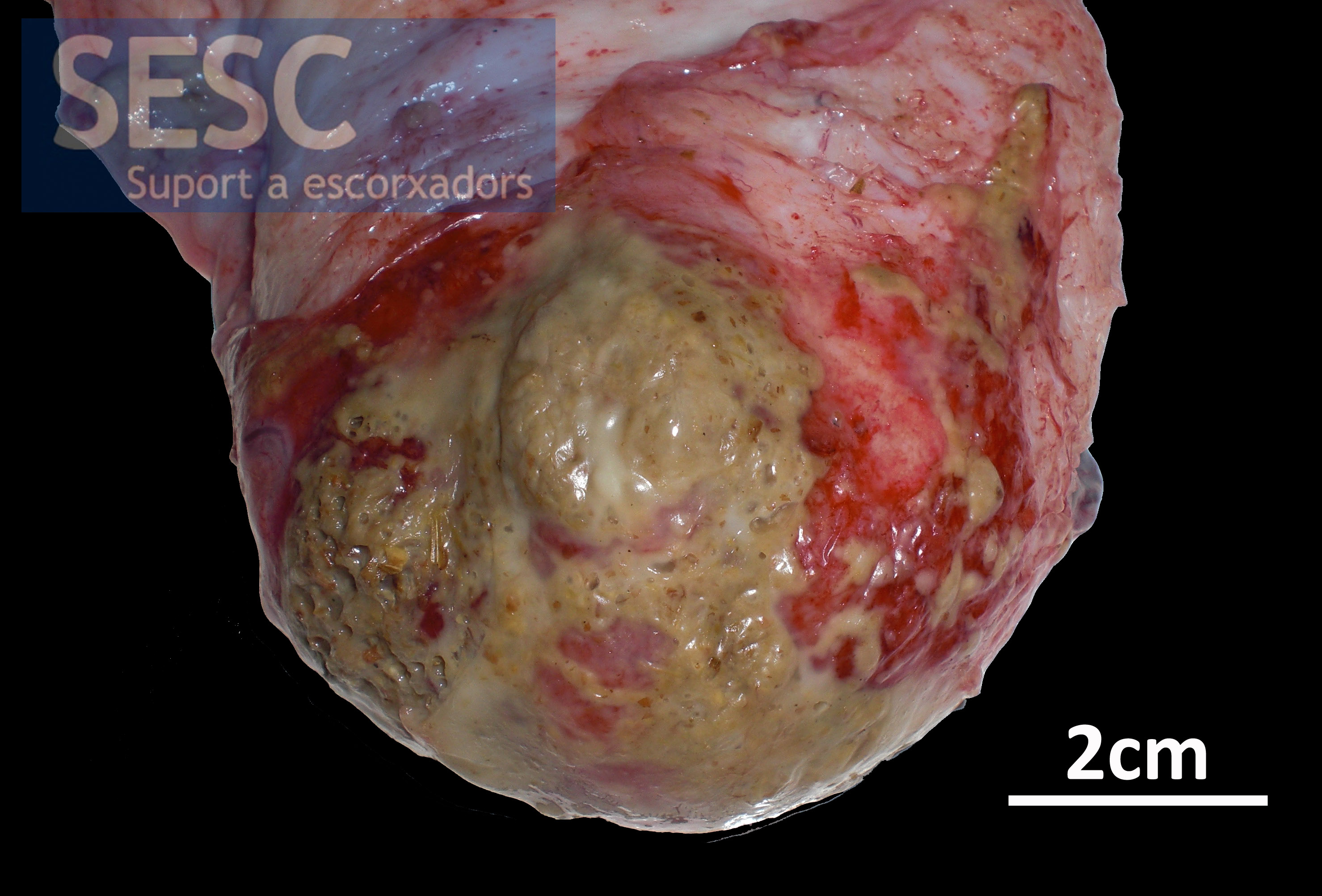
Detail of the urinary bladder aspect.

Microphotograph evidencing remains of vegetal material among the inflammatory cell infiltrate located in the serosa of the bladder (muc: mucosa, ser: serosa, musc: muscular).
