B cell leukemia in a pig carcass
In a male, cross-breed, 6 months old pig carcass the inspectors observed a change in the color of the vertebrae and sternum accompanied by a loss of consistency of the bone marrow.
The histopathological study evidenced a neoplastic proliferation of round cells that occupied almost all the space in the bone marrow.
The immunopositivity of neoplastic cells to CD20 marker is indicative of a B cell population. This marker is not present in plasma cells, so we can rule out a plasmacitoma or a myeloma. The most appropriate diagnosis in this case is a B cell leukemia. Lymphoma is discarded because no tumors were observed in other organs or extramedular infiltration. So we understand that the process is restricted to the bone marrow and, most likely, to the blood.
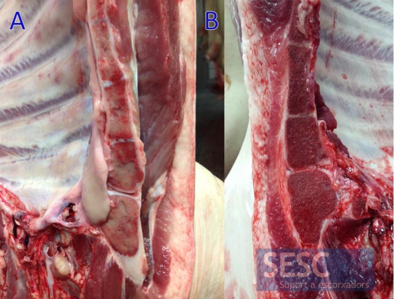
The sternum bone marrow showed a pale color (A) compared to a healthy animal (B).
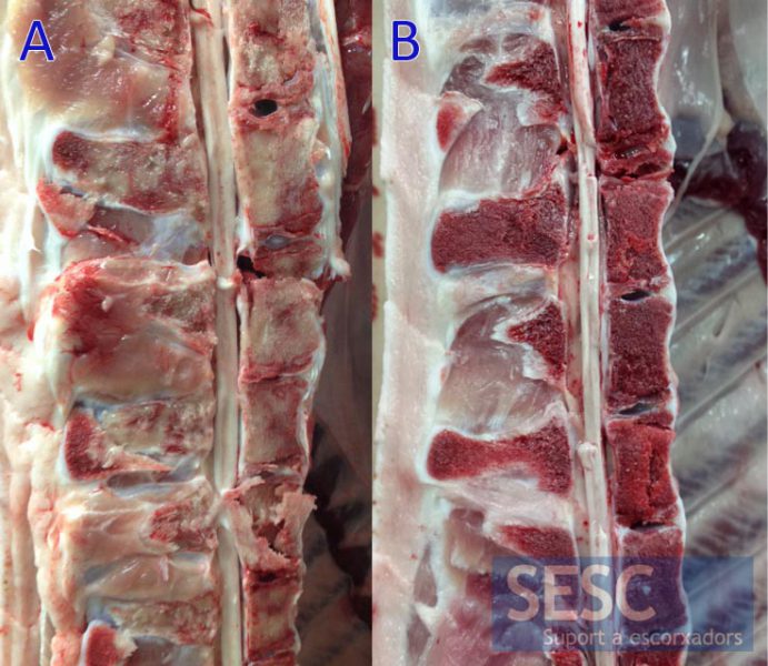
This alteration was also appreciated on vertebrae (A) compared to a healthy animal (B).
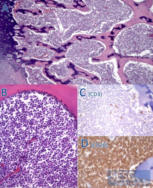
Histologically there was a neoplastic proliferation that occupied the entire bone marrow (A). At higher magnification it could be observed that these were round cells of lymphoid origin (B) which, by immunohistochemistry, were mostly identified as B cells due to the positivity against CD20 (D) and negativity against CD3, a T-cell marker (C).
