13/06/2016
|
Abdominal cavity (porcine)
1
What is your diagnosis? (31)
Time to test your microbiological skills. What do you think was cultured form these nodules? At the end of the post you can vote for the right diagnosis!
In a female, cross-bred, 3 years old pig carcass 2 well-encapsulated nodules were observed, both contained caseous material organised forming concentric laminae.
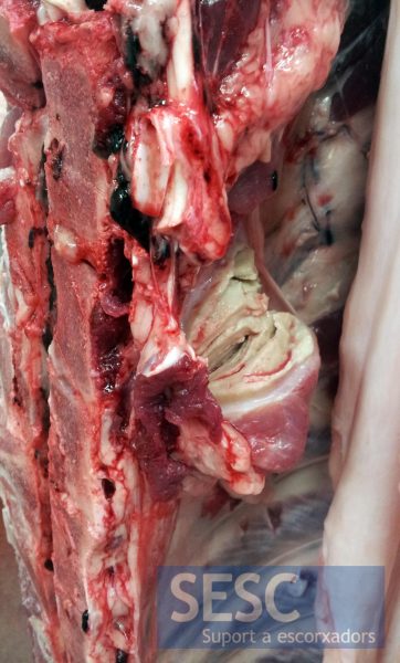
Encapsulated nodule, ventral to the vertebral column.
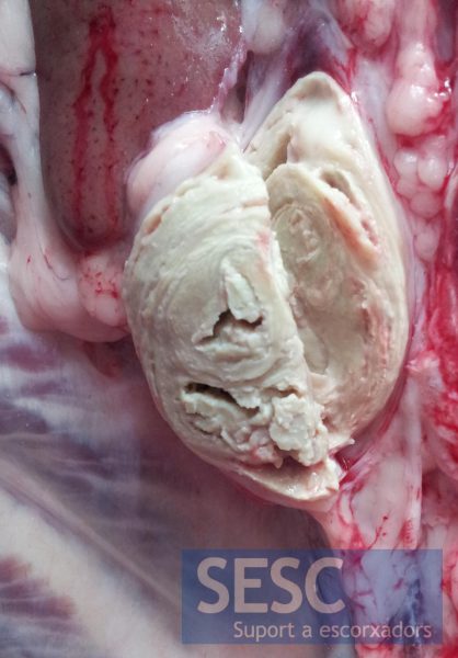
Within the nodules, caseous material is found, arragend in concentric laminae.


1 comment(s)
Comment from the LinkedIn group Veterinary pathology by Larry Kwame Tay:
I also suspected Mycobacterium sp. until I saw Corynebacterium pseudotuberculosis as part of the options. It turn out Actinomyces pyogenes rather.Pretty nice case..For most of us, Mycobacterium sp. comes to mind first when it is a case of granulomatous inflammation.